Figure 4. PT9 had an autosomal recessive bestrophinopathy (ARB). A: The fundus photograph depicts scattered subretinal yellowish lesions without classic vitelliform lesions. B: Fundus autofluorescence shows scattered hypo-autofluorescent punctate changes in the macular region and mid-periphery. C: The macular OCT image shows a schisis-like cystoid macular edema with fragmentation of the ellipsoid zone and RPE. D: The full field flash electroretinogram demonstrates a rod>cone pattern of retinal dysfunction. E: The electrooculogram shows an Arden ratio well below 1.5. All these features have been previously reported in patients with
ARB. After a nonsense BEST1 mutation was found initially via NGS sequencing, follow-up duplication/deletion testing revealed an exon 1–2 deletion in
the BEST1 gene, which was confirmed to be in trans using parental analysis, consistent with the diagnosis of ARB.
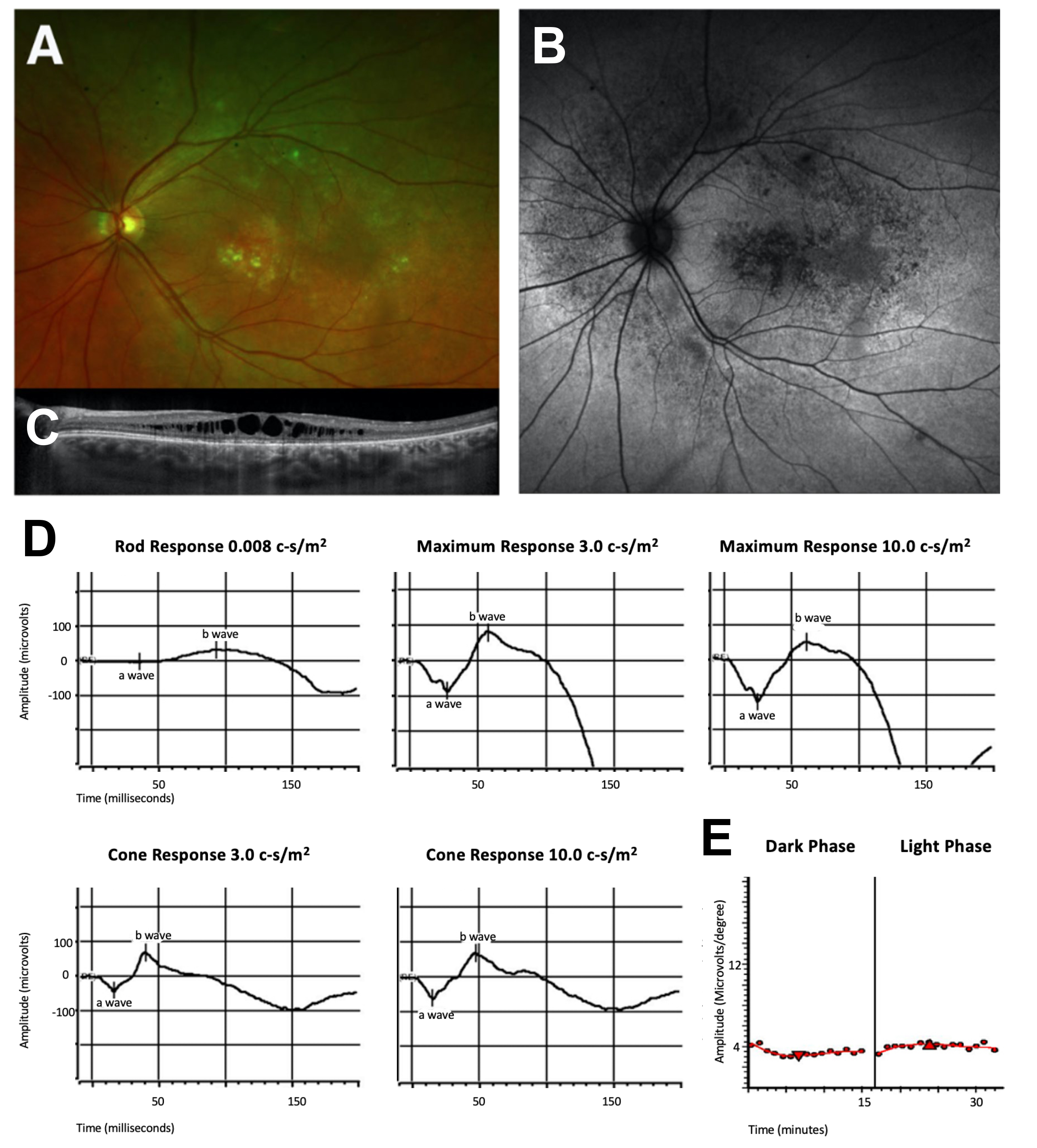
 Figure 4 of
Gupta, Mol Vis 2022; 28:203-219.
Figure 4 of
Gupta, Mol Vis 2022; 28:203-219.