Figure 6. Localization of β-catenin and active β-catenin expressions in hsa-miR-150-5p-transfected cells in Real Architecture For 3D
Tissue (RAFT). Shown are representative confocal images of the transfected CESC primary culture cells grown in RAFT TEs immunostained
for A: β-catenin and B: active- β-catenin. Nuclei were represented in blue and protein expression in red. Compared with the control- and mimic-transfected
cells, the expression levels of β-catenin and active β-catenin were higher in the inhibitor-transfected cells with nuclear
translocation. Nuclear localization of active β-catenin indicates active Wnt signaling. Scale bar: 50 µm.
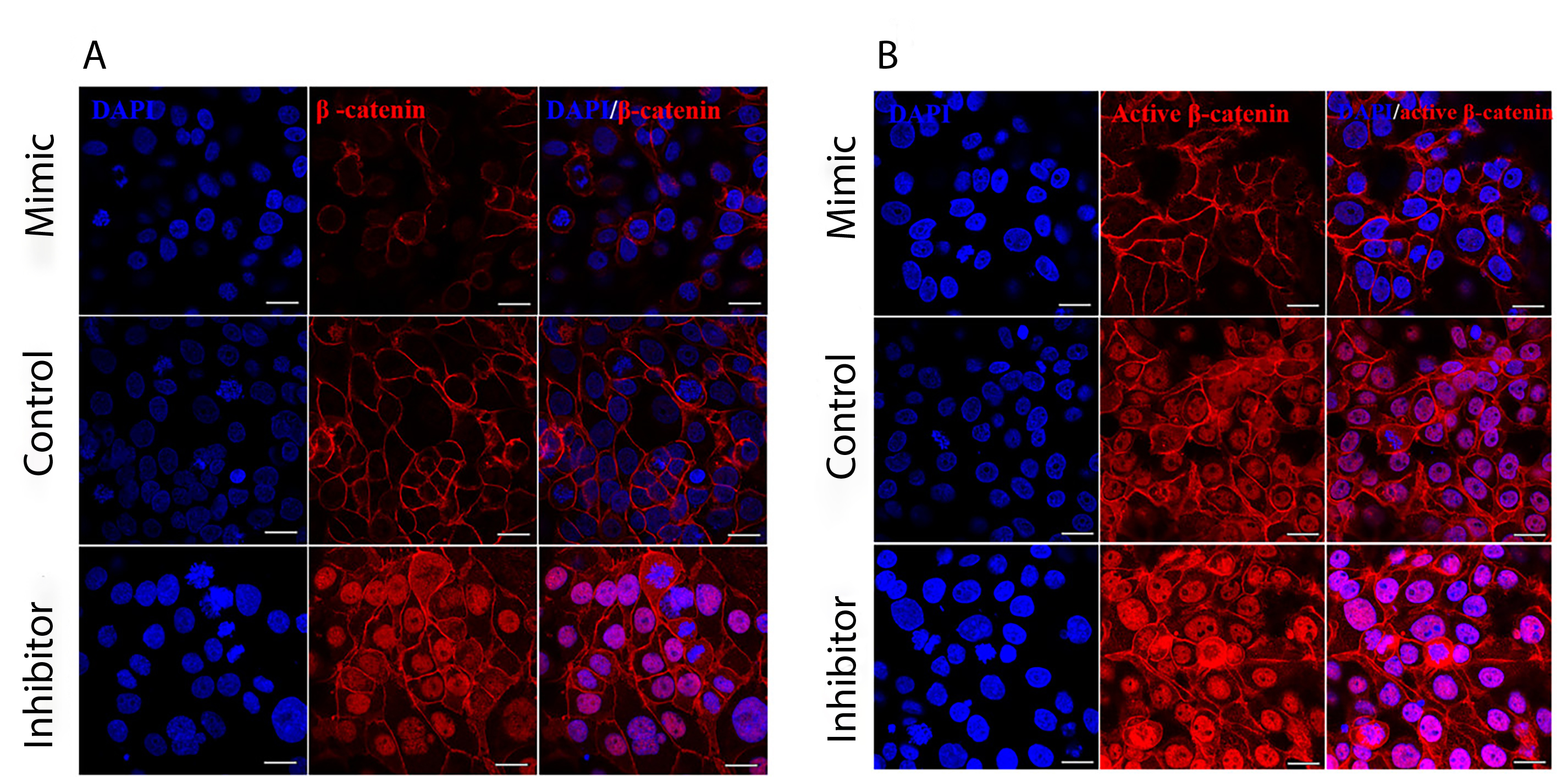
 Figure 6 of
Kalaimani, Mol Vis 2022; 28:178-191.
Figure 6 of
Kalaimani, Mol Vis 2022; 28:178-191.