Figure 3. Localization of protein expression in hsa-miR-150-5p-transfected cells. The figures present the representative confocal images
of transfected limbal epithelial cells immunostained for AKT3, JARID2, β-catenin, active β-catenin, AXIN2, ABCG2, p63⍺, and
Cx43. Nuclei were represented in blue and protein expression in green or red. The cells with nuclear positivity were marked
with asterisks in the Wnt signaling regulators (β-catenin, active β-catenin, and AXIN2). Nuclear localization of active β-catenin
is the indication of active Wnt signaling. Scale bar: 50 µm.
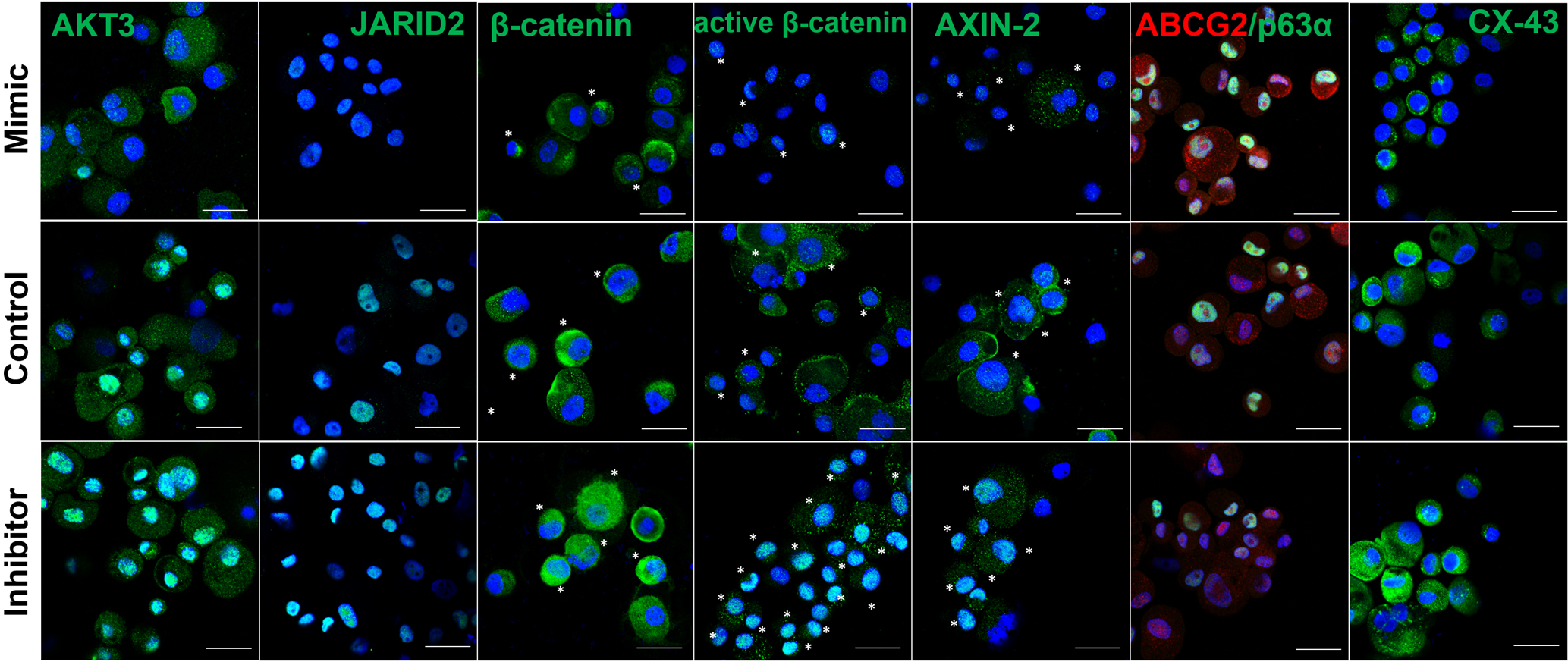
 Figure 3 of
Kalaimani, Mol Vis 2022; 28:178-191.
Figure 3 of
Kalaimani, Mol Vis 2022; 28:178-191.