Figure 2. Protein expression profile of the hsa-miR-150-5p-transfected cells grown in the two-dimensional culture system. A: The representative western blots of the proteins of interest (AKT3, p-AKT3, JARID2, β-catenin, active β-catenin [ABC], AXIN2,
ABCG2, ΔNp63⍺, and Cx43) in the three groups (hsa-miR-150-5p mimic, inhibitor, and control, n = 3) are shown. GAPDH was used
as a normalizing reference and loading control. B: The relative expression profiles of the proteins were quantified by western blotting. *p < 0.05; **p < 0.001; ***p < 0.0001.
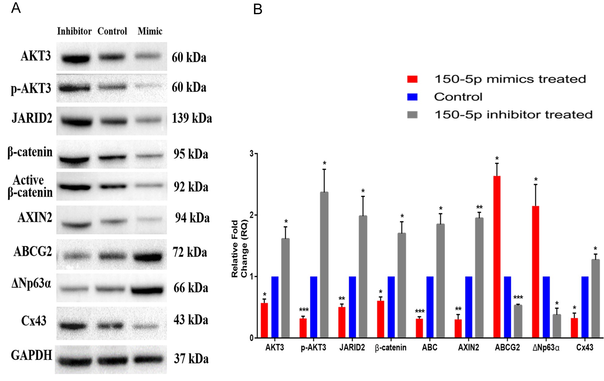
 Figure 2 of
Kalaimani, Mol Vis 2022; 28:178-191.
Figure 2 of
Kalaimani, Mol Vis 2022; 28:178-191.