Figure 4. Integrity of optic nerve axons following intraocular pressure (IOP) elevation with or without macitentan treatment compared
to naive animals. Following 4 weeks of IOP elevation, rats were euthanized, and optic nerve sections obtained were subjected
to PPD staining to assess optic nerve degeneration. Axonal degeneration accompanied by gliosis and glial scar were observed
in IOP-elevated untreated rats compared to naïve eyes. IOP-elevated macitentan-treated rats show significant protection of
their axons, compared to those of untreated rats. Pink arrows point to glial scarring, which was found mainly in the retinas
from IOP-elevated untreated rats. Dark spots (yellow arrowheads) indicate the collapsed axons in the untreated IOP-elevated
rats (A). The mean counts of healthy axons were significantly reduced in untreated IOP-elevated animals compared to naïve animals
(*p=0.022, n=7) and a significant protection was seen in macitentan-treated rats with IOP elevation (*p=0.013, n=7; B) compared to untreated IOP-elevated rats (one-way ANOVA followed by Tukey’s multiple comparisons test). Scale bar: 20 μm.
n=7 (3 male rats and 4 female rats) for untreated rats, n=7 (3 male rats and 4 female rats) for macitentan-treated rats and
n=10 (5 male rats and 5 female rats) for naïve rats.
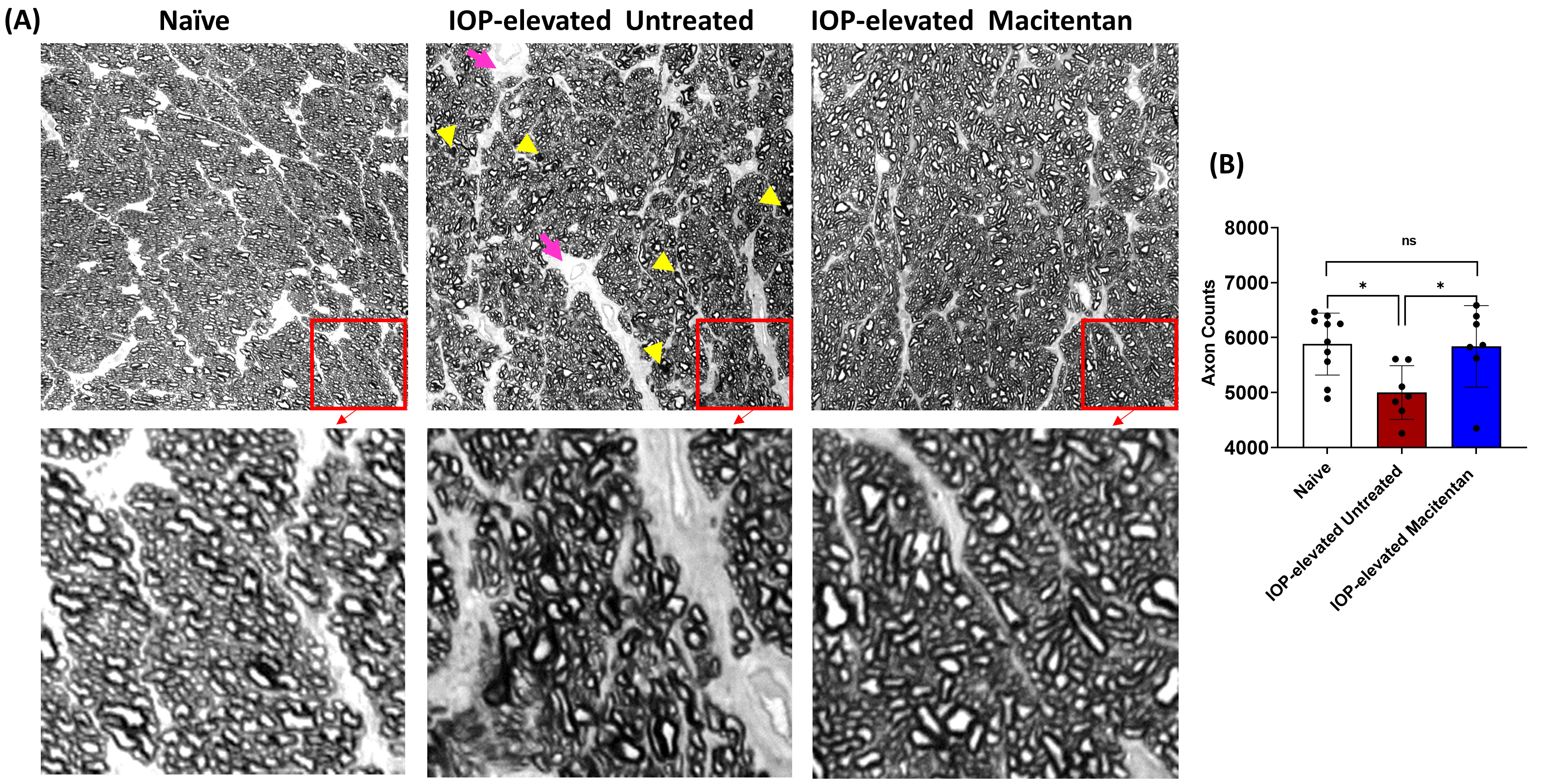
 Figure 4 of
Kodati, Mol Vis 2022; 28:165-177.
Figure 4 of
Kodati, Mol Vis 2022; 28:165-177.