Figure 2. Macitentan preserves retinal ganglion cell (RGC) function in Brown Norway rats following intraocular pressure (IOP) elevation.
Pattern electroretinography (ERG) measurements in naive, IOP-elevated untreated, and IOP-elevated macitentan-treated retired
breeder Brown Norway rats (9 to 13 months old) at 2 weeks and 4 weeks following IOP elevation by the Morrison method. A significant
loss of pattern ERG (PERG) amplitude was observed during 2 weeks (A) and 4 weeks (B) of IOP elevation in untreated rats compared to naive animals. A protective trend against the decline in PERG amplitude was
found in macitentan-treated rats compared to untreated IOP-elevated rats at 2 weeks (A) and 4 weeks (B). PERG waveforms for naïve, IOP-elevated untreated and IOP-elevated macitentan-treated retired breeder Brown Norway rats are
shown in panel (C). PERG waveform obtained consisted of a major peak (P1) followed by a trough (N2). PERG amplitude was measured from peak-to
trough (P1 to N2) of the waveform, while PERG latency was the time needed to attain the P1 peak of the waveform (C). *p<0.05, **p<0.005 indicates statistical significance, one-way ANOVA, followed by Tukey’s multiple comparison test, n=6
(3 male rats and 3 female rats) for untreated rats, n=7 (4 male rats and 3 female rats) for macitentan-treated rats, and n=10
(5 male rats and 5 female rats) for naïve rats.
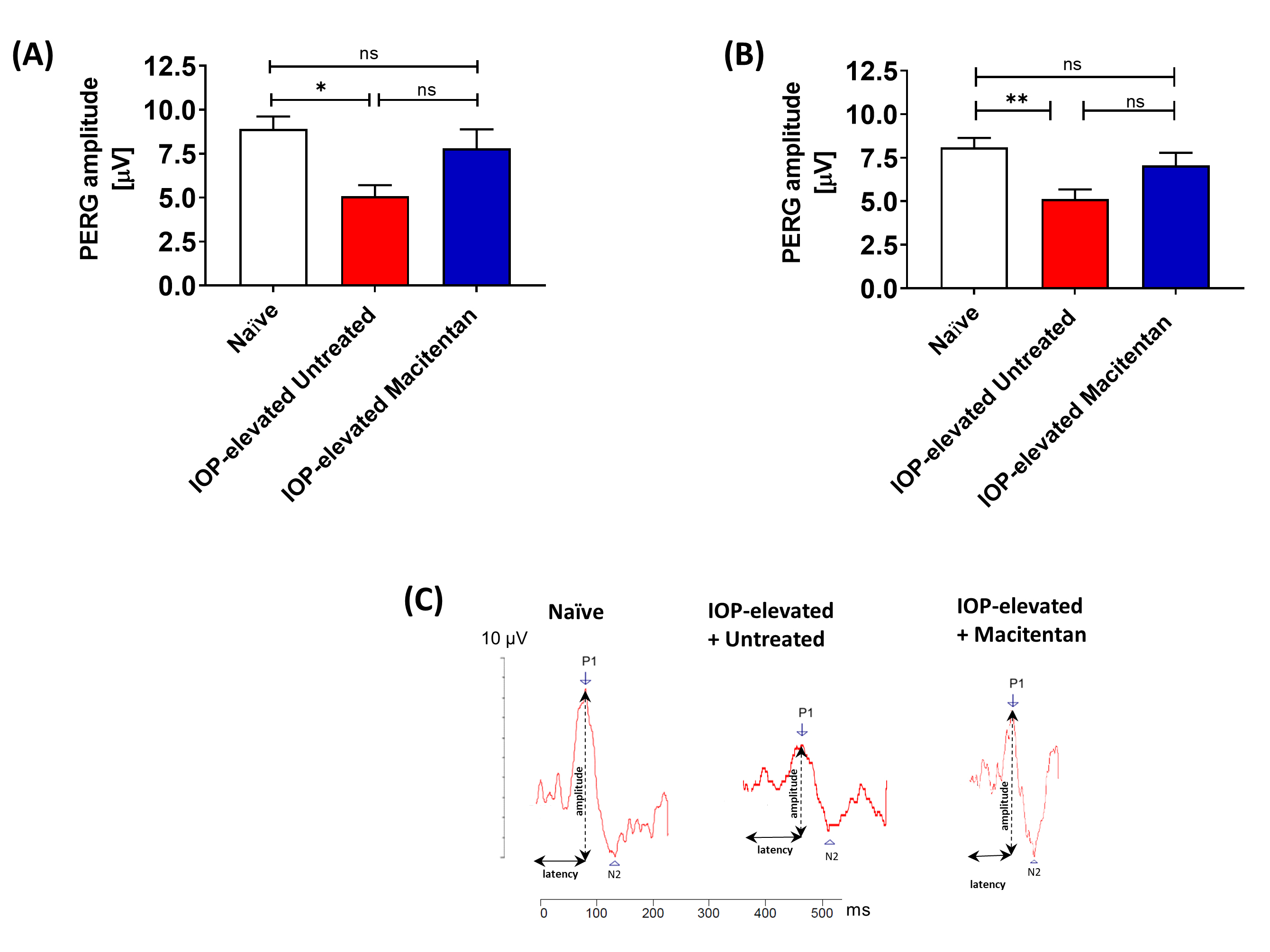
 Figure 2 of
Kodati, Mol Vis 2022; 28:165-177.
Figure 2 of
Kodati, Mol Vis 2022; 28:165-177.