Figure 6. Determination of the effects of the αAP1 and αBP4 peptides on the secondary structure of αA- and αB-crystallins with CD spectroscopy.
For circular dichroism (CD) spectral analysis, 10 µM protein (αA- or αB-crystallin) and 25 µM peptide (αAP1 or αBP4) in 200
µl reaction mixtures of 10 mM phosphate buffer (pH 7.4) were incubated at 37 °C for 15 min, and the secondary structures were
recorded in the far-ultraviolet (UV) region (190–260 nm) using a Jasco CD spectrometer and appropriate controls (αA- or αB-crystallin
alone and the αAP1 or αBP4 peptide alone). The spectrum of the buffer alone was subtracted from each spectrum, and the secondary
structure was predicted using the Selcon 3 program.
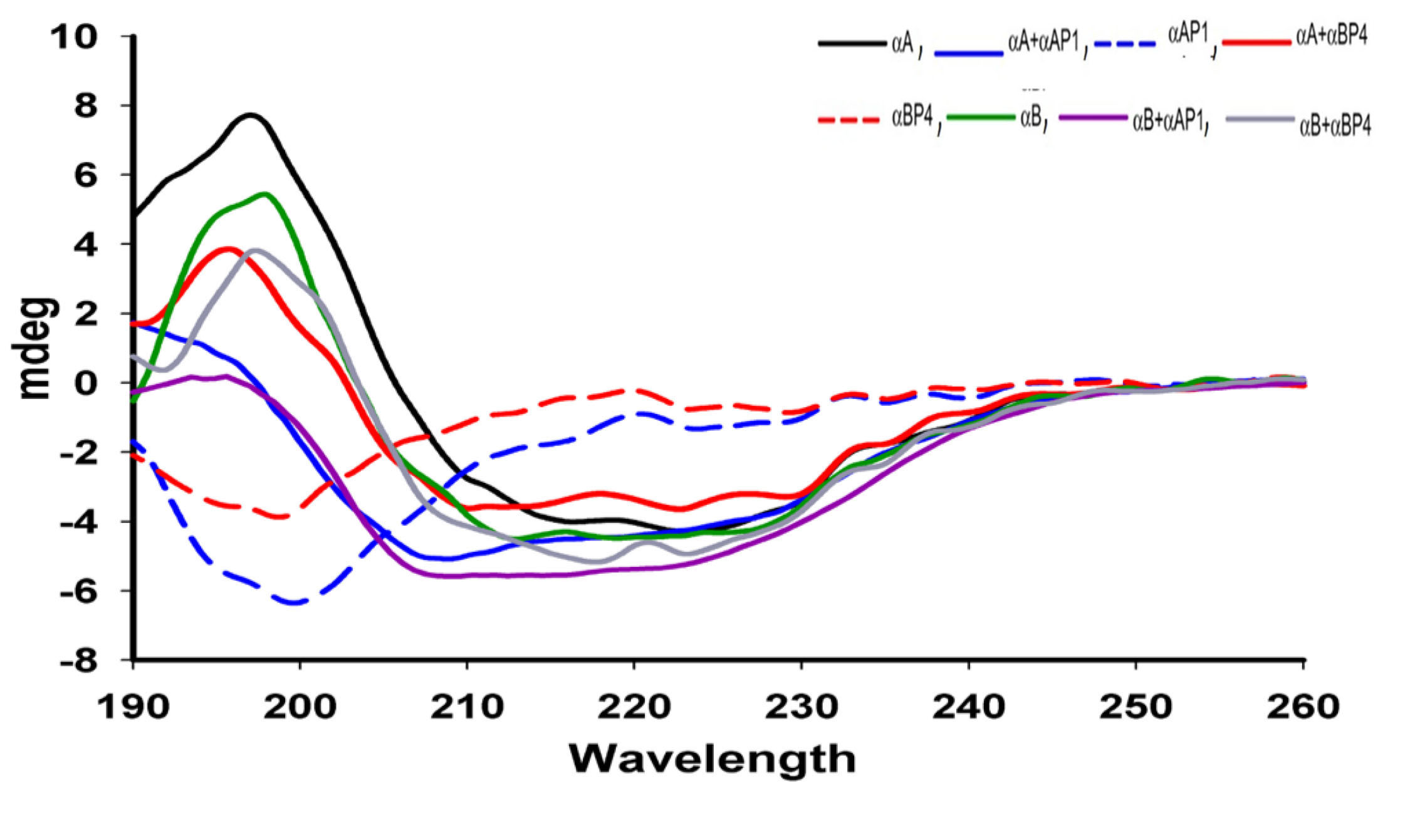
 Figure 6 of
Srivastava, Mol Vis 2022; 28:147-164.
Figure 6 of
Srivastava, Mol Vis 2022; 28:147-164.