Figure 2. Diquafosol tetasodium-induced expression of mucins in hyperosmotic stressed HCECs. A: Concentrations of secreted MUC5AC by ELISA before and after hyperosmotic stress (400 mOsm/l) with diquafosol tetrasodium.
B: Gene expression of MUC5AC before and after hyperosmotic stress with diquafosol tetrasodium. C: Transcription of membrane-associated mucin MUC1 and MUC16 genes in HCECs induced by diquafosol tetrasodium. MUC1 and MUC16
mRNA levels are shown. D: MTT cell proliferation/cytotoxicity assay of HCECs with various concentrations of diquafosol tetrasodium. E: Representative confocal images of HCECs immunostained for MUC1, MUC5AC, and MUC 16. Bar = 20 μm. The values in these graphs
represent the mean of three experiments; the error bars represent the standard deviation. * p<0.05, ** p<0.01.
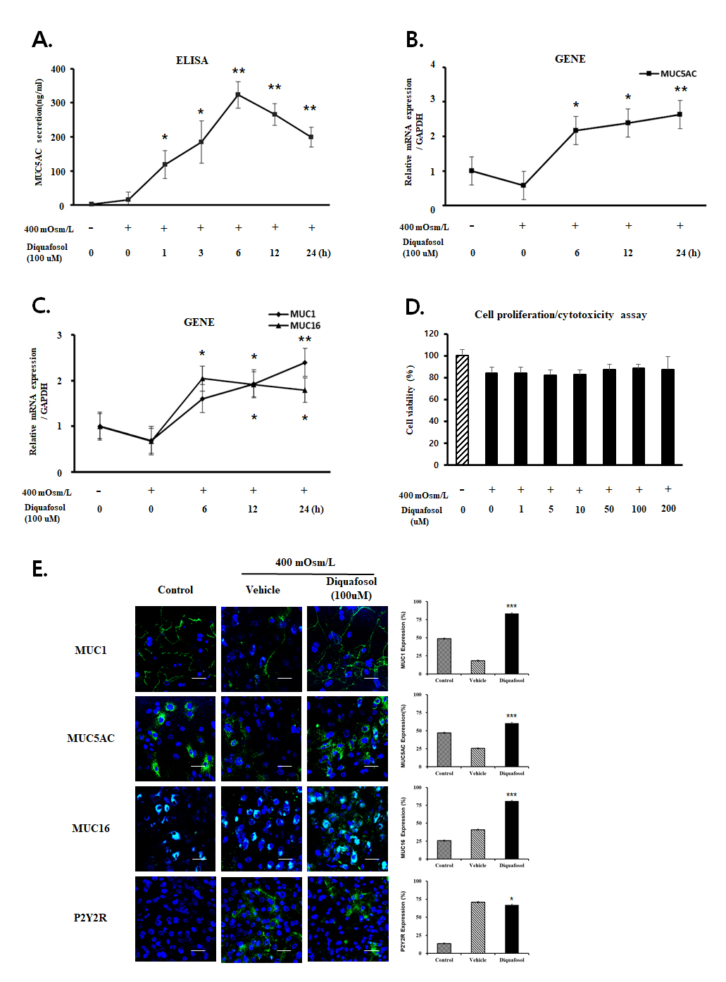
 Figure 2 of
Lee, Mol Vis 2022; 28:114-123.
Figure 2 of
Lee, Mol Vis 2022; 28:114-123.