Figure 1. Flow diagram for the production of human conjunctival epithelial cells from cadavers. Conjunctival specimens from cadavers
were incubated at 4 °C with 0.1% protease. Suspended epithelial cells were seeded in culture dishes. When these cells reached
60%–70% confluence, they were harvested and seeded onto transwell inserts using 105 cells per insert in culture media. At confluence, the medium was removed to establish an air-liquid interface (ALI) culture
to promote differentiation and stratification. After airlift culturing for two weeks to induce multi-layer human conjunctival
epithelial cells (HCECs), hyperosmotic stress (400 mOsm/l) was applied by adding an appropriate volume of 5 M NaCl to ALI
media with diquafosol tetrasodium. Immunofluorescence microscopy shows multi-layered HCECs stained with CK19 shown in green.
Bar = 50 μm.
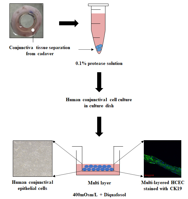
 Figure 1 of
Lee, Mol Vis 2022; 28:114-123.
Figure 1 of
Lee, Mol Vis 2022; 28:114-123.