Figure 3. The clinical features of the individuals with LCA. A: Patient MEP-305. B: Patient MEP-318. The top row shows fundus imaging. The second row shows autofluorescence (AF) imaging. The third row shows
optical coherence tomography (OCT) imaging. The bottom row shows full-field electroretinography (ffERG). In patient A, there
are vascular attenuation, RPE atrophy with increased visibility of choroidal vessels, and fine granular pigmentation just
outside the vessels. The pigmentary changes inferiorly are secondary to laser for a Coats-like reaction that the patient developed.
AF imaging showed a perimacular hyper-AF ring. In patient B, the fundus of both eyes indicates moderate waxy pallor, mottling
in macula, as well as moderate vascular attenuation. AF imaging shows peripheral hypo-AF and hyper-AF rings of the parafovea
and midperiphery OU. The ffERG of patients A and B reveals severe cone and rod dysfunction. OCT imaging shows that the two
patients have a relatively normal foveal structure. C–E: Phenotypes of patient RKK_665. C: Retinal photographs of both eyes illustrating optic disc pallor and diffuse retinal pigmentation and atrophy with arteriolar
narrowing but much more pronounced inferiorly in both eyes. This corresponds to the absence of Goldmann visual fields superiorly
OU. In the periphery, there are marked nummular pigmented clumps and areas of atrophy. D: Thinning of the retina and a small remaining subfoveal ellipsoid zone (EZ), illustrating the remaining photoreceptors, likely
cones. E: Significant remaining central and inferior field in both eyes. The superior visual fields are absent in both eyes.
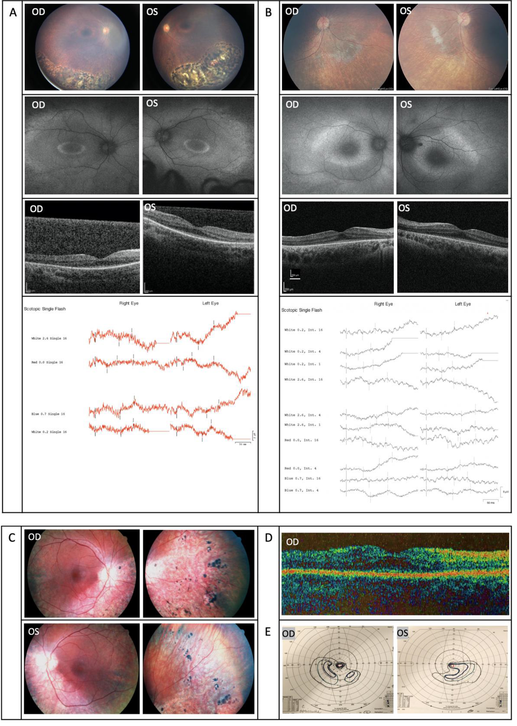
 Figure 3 of
Zou, Mol Vis 2021; 27:95-106.
Figure 3 of
Zou, Mol Vis 2021; 27:95-106.