Figure 8. Fundus autofluorescence and OCT imaging. The images are referred to patient 2 (A) and patient 1 (B). The yellow line indicates the vertical OCT scan line passing through the hyper-AF demarcation line while the blue line
indicates the intact EZ band. The two white arrows indicate the internal and external borders of the transition zone.
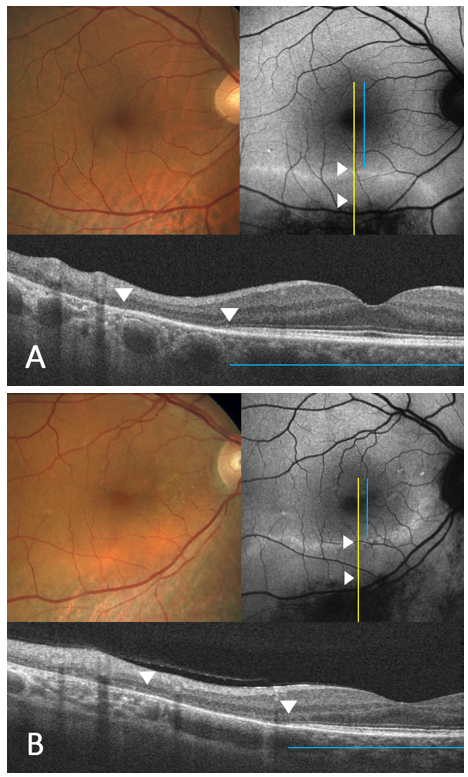
 Figure 8 of
Verdina, Mol Vis 2021; 27:78-94.
Figure 8 of
Verdina, Mol Vis 2021; 27:78-94.