Figure 3 of
Verdina, Mol Vis 2021; 27:78-94.
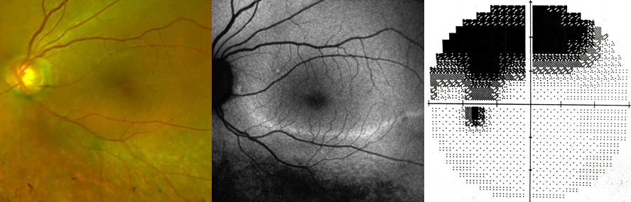
Figure 3.
Color retinography, fundus autofluorescence, and visual field testing for left eye in patient 1. The image shows the correspondence between the inferior retinal degeneration and the superior hemifield defect.
 Figure 3 of
Verdina, Mol Vis 2021; 27:78-94.
Figure 3 of
Verdina, Mol Vis 2021; 27:78-94.