Figure 1 of
Altay, Mol Vis 2021; 27:757-767.
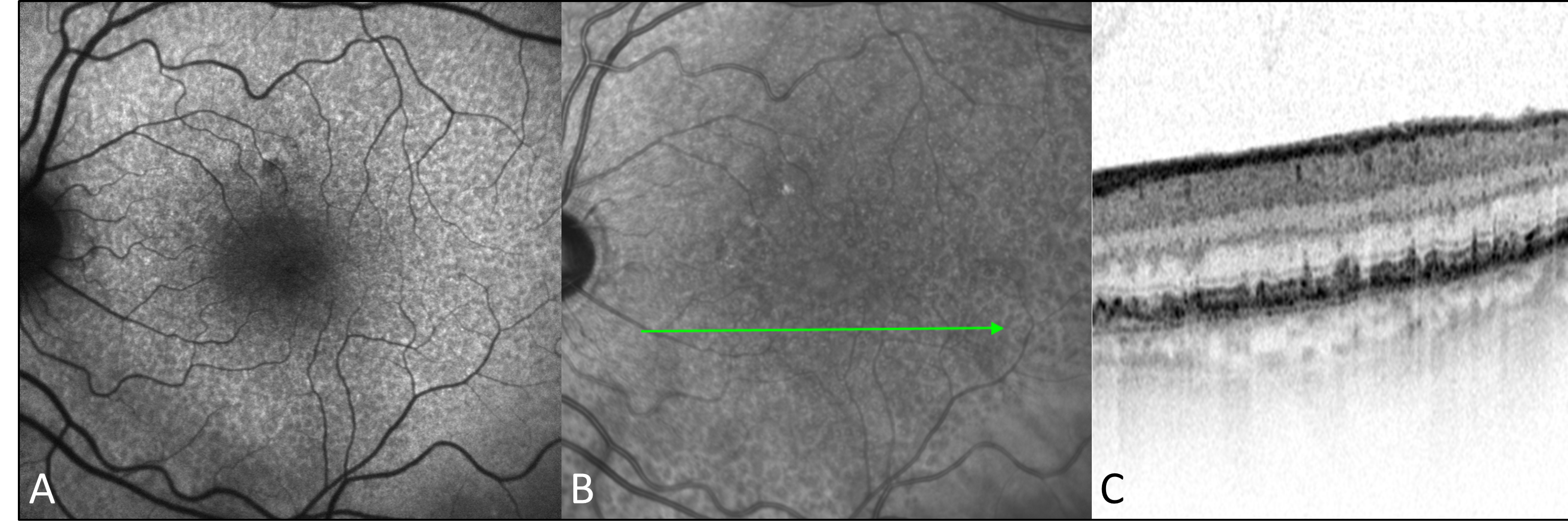
Figure 1.
Imaging of reticular pseudodrusen. Example of reticular pseudodrusen (RPD) visible via fundus autofluorescence (
A
), infrared imaging (
B
) and spectral-domain optical coherence tomography (SD-OCT;
C
).
 Figure 1 of
Altay, Mol Vis 2021; 27:757-767. Figure 1 of
Altay, Mol Vis 2021; 27:757-767.
Figure 1 of
Altay, Mol Vis 2021; 27:757-767. Figure 1 of
Altay, Mol Vis 2021; 27:757-767.