Figure 5. Localization of the macrophage marker F4/80 to clump cells in type 5 adenovirus (Ad5)-injected eyes. Fluorescent and light
micrographs of eyes immunostained with F4/80 and counterstained with 4’,6-diamidino-2-phenylindole (DAPI; nuclear stain) in
treated eyes of mice from the Saline cohort (left column) compared with those of mice from the Virus cohort (right column).
A-B: The central cornea shows a similar localization and prevalence of F4/80+ cells. C-F: the mid-peripheral iris and G-J: iridocorneal angle show an increased prevalence of F4/80+ cells localized along the anterior iris stroma, matching the location of clump cells visualized via slit-lamp exam (white arrowheads). Note that F4/80+ cells also appear to be pigment laden, which is an additional feature ascribed to clump cells. F4/80+ cells are also prominent in the posterior iris pigmented epithelium and iridocorneal angle of Ad5-injected eyes. Notation
of the prominent intraocular structures is indicated (white text) in panel G. Scale bar = 100 µm for (A-F) and (G-J).
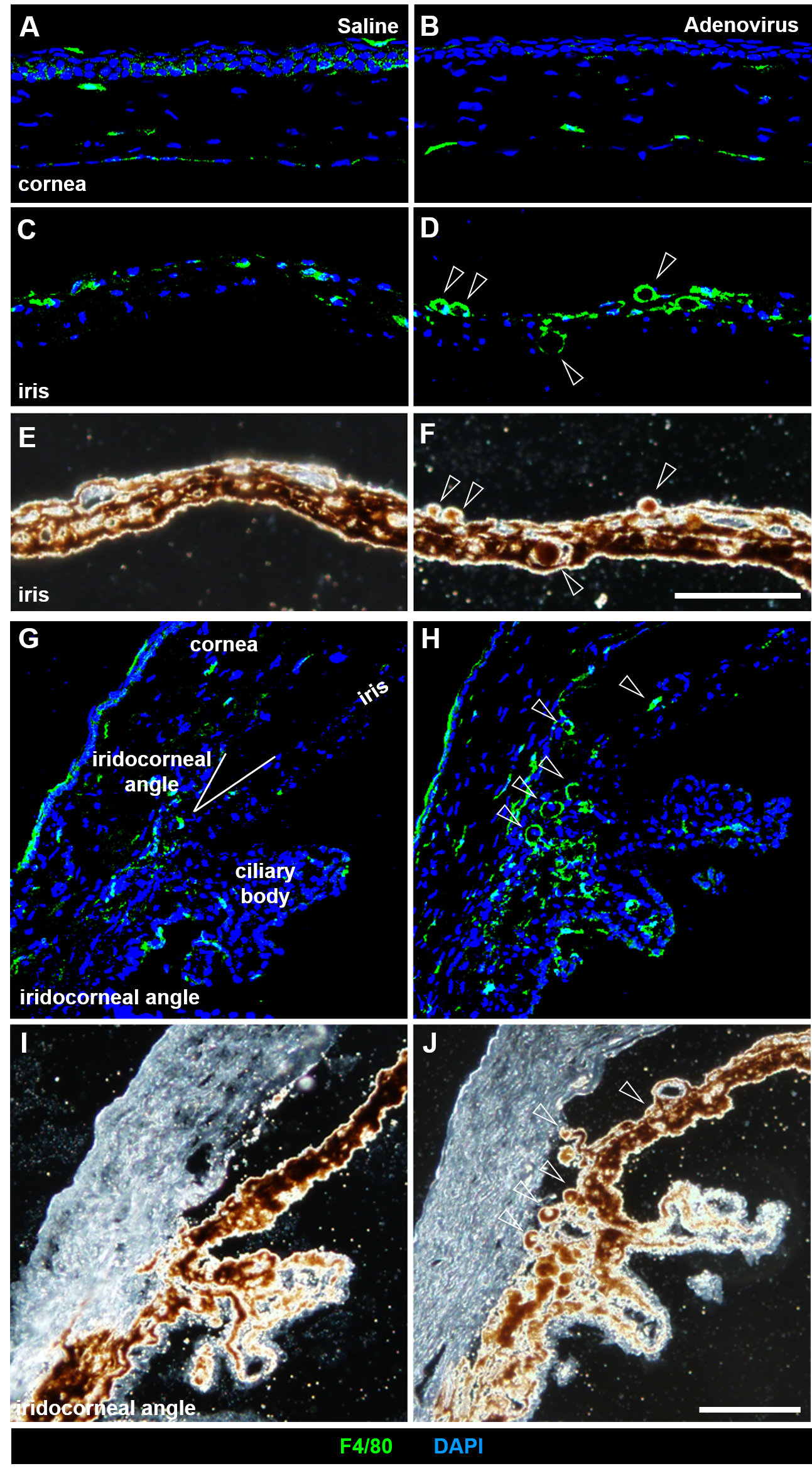
 Figure 5 of
Meyer, Mol Vis 2021; 27:741-756.
Figure 5 of
Meyer, Mol Vis 2021; 27:741-756.