 Appendix 9 of
Meyer, Mol Vis 2021; 27:741-756.
Appendix 9 of
Meyer, Mol Vis 2021; 27:741-756. Appendix 9 of
Meyer, Mol Vis 2021; 27:741-756.
Appendix 9.
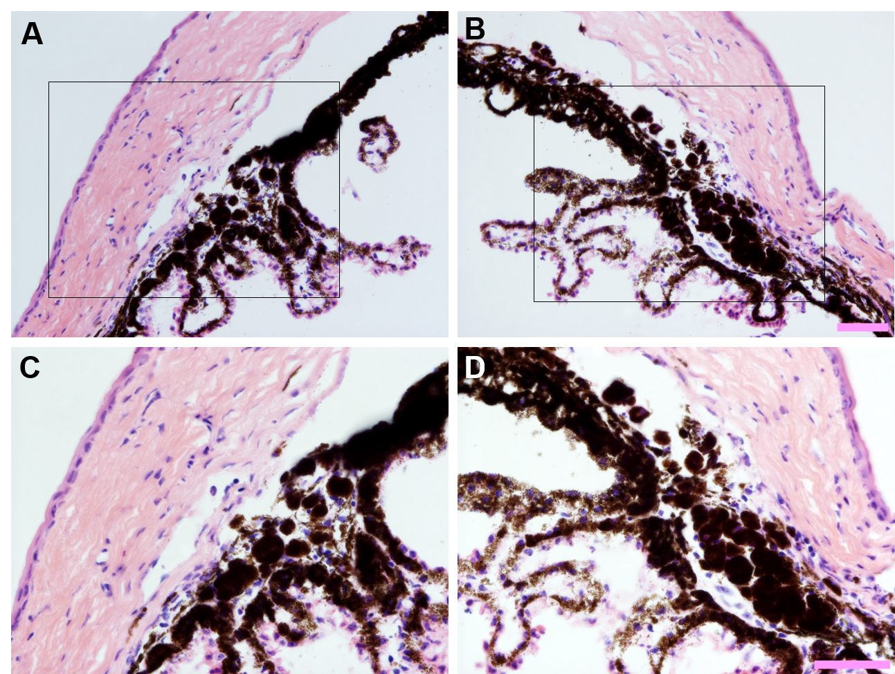
To access the data, click or select the words “Appendix 9.” Persistence of clump cells in the iridocorneal angle of eyes following intraocular Ad5 injection. Light micrographs collected from opposite poles (left vs. right column) of hematoxylin and eosin-stained histological sections from the same eye shown at (A–B) 200X and (C–D) 400X total magnification. Areas within inset boxes (top row) are show at higher magnification below (bottom row). Note that despite some variability, there is a persistent localization of clump cells deep within the iridocorneal angle with proximity to the drainage structures. Scale bar = 50 µm.