Figure 3. Transmission electron micrographs of RGCs in the GCL and inner plexiform layer (IPL). Representative ultrastructural images
show the presence of double- or multiple-membrane autophagic vesicles (box) containing cell organelles in the cytoplasma of
ganglion cell layer (GCL) retinal ganglion cells (RGCs) and dendrites of IPL RGCs of sham-operated (Control) rats or EVC-operated
rats treated with vehicle (Operated), TMP (TMP), TMP+Rapamycin (TMP+Rapamycin), or TMP+Ly294002 (TMP+Ly294002). Three eyes
were examined in each experimental period.
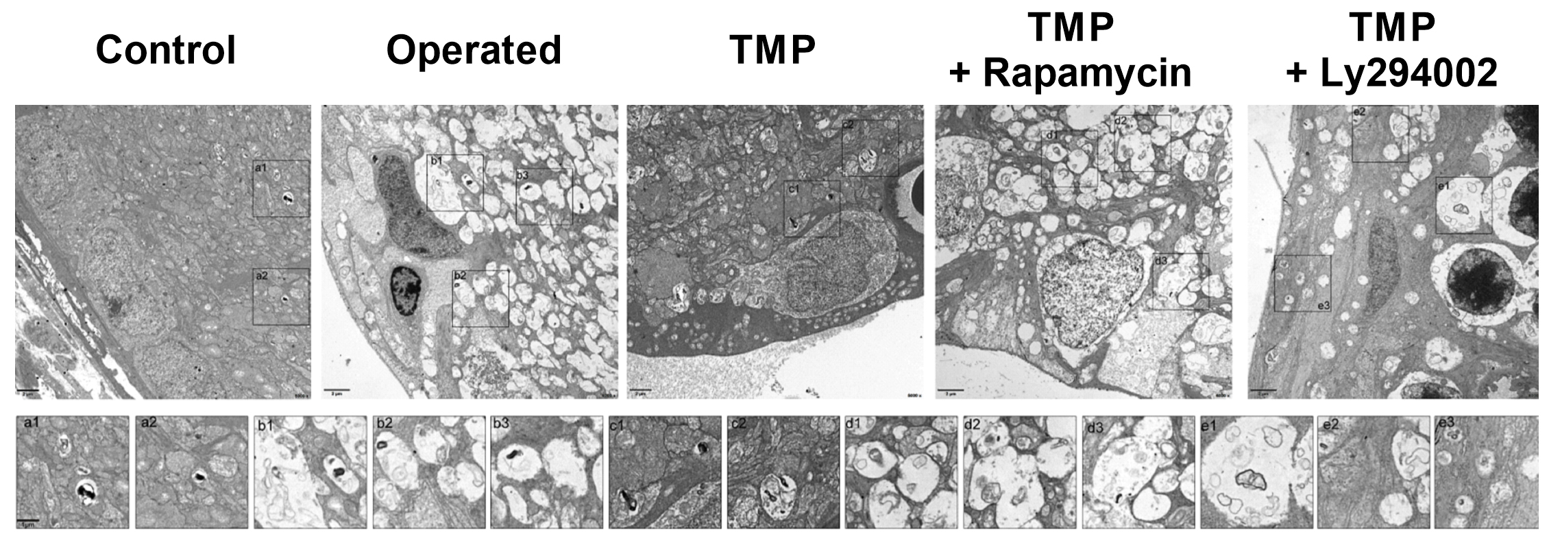
 Figure 3 of
Du, Mol Vis 2021; 27:725-733.
Figure 3 of
Du, Mol Vis 2021; 27:725-733.