Figure 4. Simulated three-dimensional crystal structures of proteins. Predicted crystal structures of wild-type (left) and mutant (right)
proteins. The wild-type (left) and mutant (right) residues are green, while the residues that bind with them are yellow (A–H).
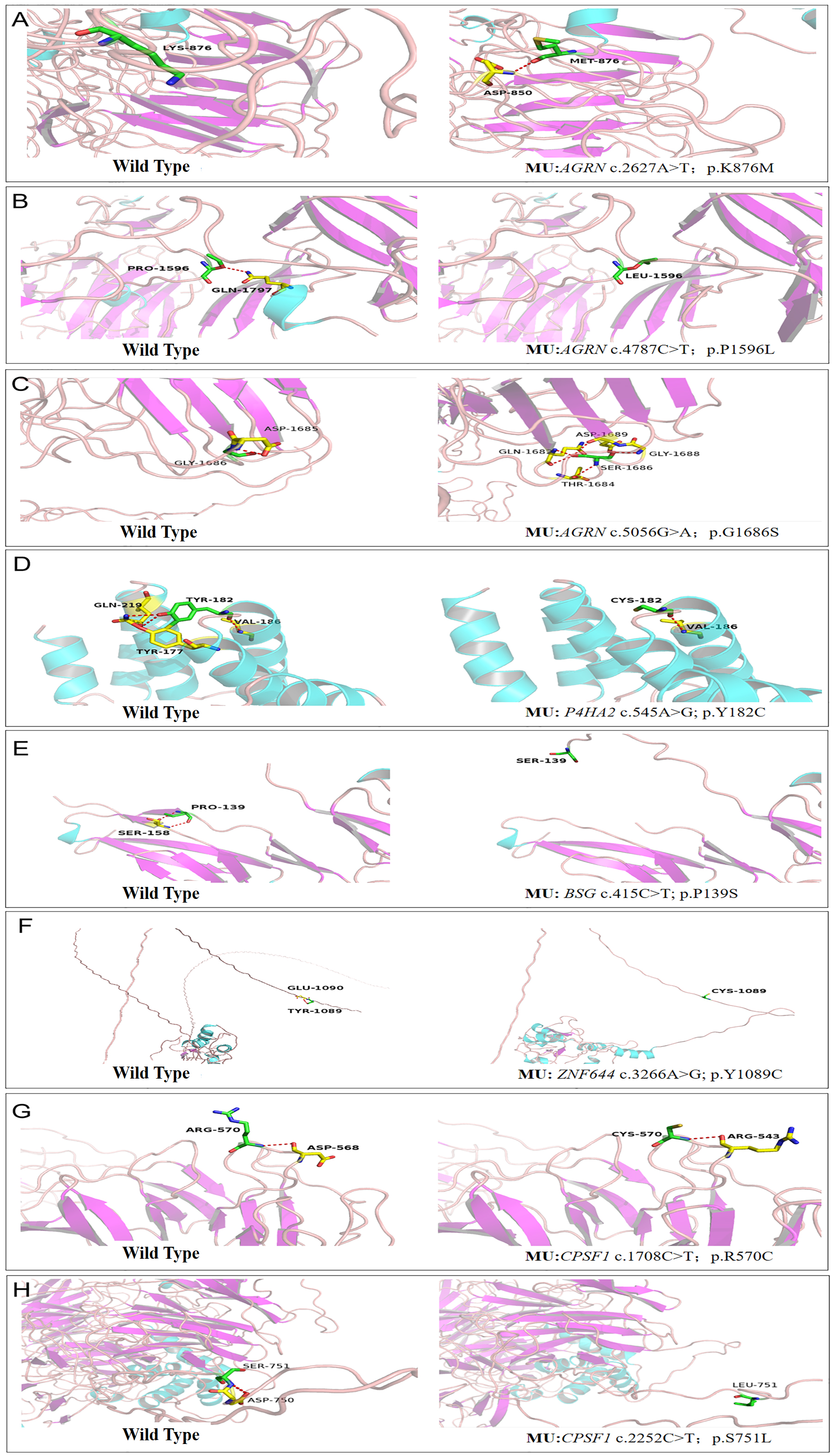
 Figure 4 of
Zheng, Mol Vis 2021; 27:706-717.
Figure 4 of
Zheng, Mol Vis 2021; 27:706-717.