Figure 8. Protein treatment did not affect outflow facility in Mmp-9KO mouse eyes. A: Intraocular pressure in Mmp-9KO and C57BL/6 mouse eyes. Intraocular pressure (mean ± SD mmHg) in Mmp-9KO mouse eyes (n = 20) was significantly higher than that of C57BL/6J animals (n = 20; **** p<0.0001, t test). B: Outflow facility in Mmp-9KO and C57BL/6 mouse eyes. Outflow facility (mean ± SD µl/min/mmHg) in Mmp-9KO mouse eyes (n = 14) was significantly lower than that of C57BL/6J animals (n = 16; **** p<0.0001, t test). C: Outflow facility in BSA-, tPA-, and NE-tPA-treated Mmp-9KO mouse eyes. Outflow facility (mean ± SD µl/min/mmHg) was not significantly different across groups: Mmp-9KO+BSA (n = 21), Mmp-9KO+tPA (n = 13), and Mmp-9KO+NE-tPA (n = 8; p>0.05, ANOVA).
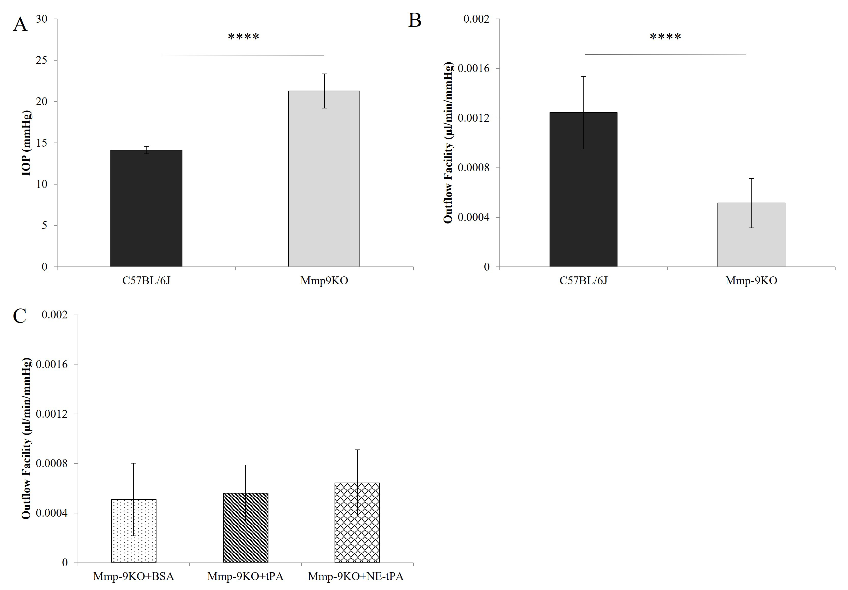
 Figure 8 of
Gindina, Mol Vis 2021; 27:691-705.
Figure 8 of
Gindina, Mol Vis 2021; 27:691-705.