Figure 3. Visualization of adenovirus expression. Flatmounts of anterior segments of mouse eyes from animals injected with (A) AdPLAT (TA+AdPLAT), (B) AdPLATNE (TA+AdPLATNE), and (C) AdNull (TA+AdNull). There was robust mCherry expression in both AdPLAT- and AdPLATNE-treated eyes (white arrowheads). There
was no mCherry expression in AdNull-treated eyes. DAPI was used as a counterstain for nuclei. Arrows indicate mCherry-positive
cells. C: qRT-PCR quantification of PLAT expression in angle ring tissues. Levels of PLAT were detected in AdPLAT (n = 16) and AdPLATNE (n = 12) eyes, but were undetectable in AdNull (n = 16) and naïve (n = 20)
eyes. TM = trabecular meshwork.
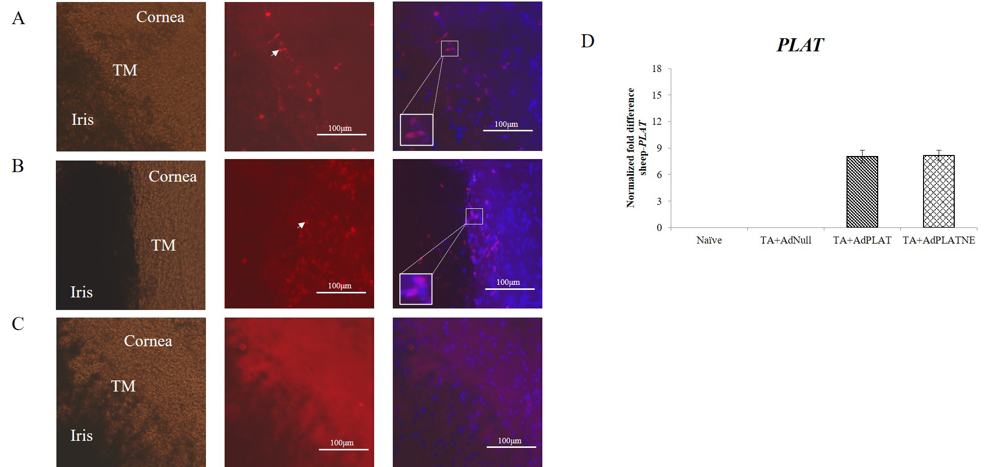
 Figure 3 of
Gindina, Mol Vis 2021; 27:691-705.
Figure 3 of
Gindina, Mol Vis 2021; 27:691-705.