Figure 5. AQP5 deficiency induced structural damage to lacrimal gland myoepithelial cells (MECs) in mice. A: Immunofluorescence staining of alpha-smooth muscle actin (α-SMA) showed myoepithelium morphology (green: α-SMA; blue: 4’6-diamidino-2-phenylindole
[DAPI]). B: ImageJ and GraphPad were used to calculate the result of A (n = 6 samples). C: Western blotting showed the expression level of α-SMA and GAPDH in lacrimal glands. D: ImageJ and GraphPad were used to compute the result of C (n = 3 samples). Data were expressed as mean ± standard deviation
(SD). *p<0.05, **p<0.001 two-way analysis of variance (ANOVA). Scale bars: (A) 10 μm, 2 μm. (D) 40 μm.
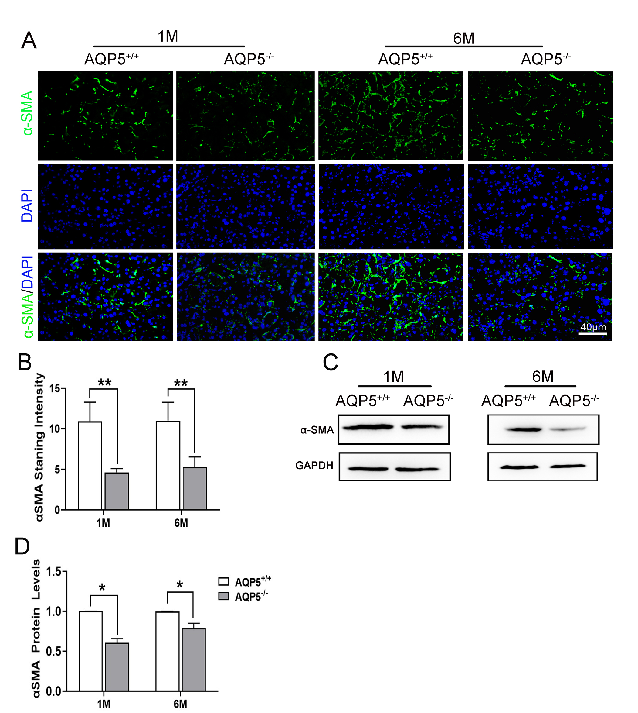
 Figure 5 of
Hu, Mol Vis 2021; 27:679-690.
Figure 5 of
Hu, Mol Vis 2021; 27:679-690.