Figure 4. AQP5 deficiency induced inflammation in the lacrimal gland. A: Hematoxylin and eosin (H&E) staining showed morphology changes. CD4+: Immunofluorescence staining of CD4+ demonstrated CD4+ T cells (red: CD4+, blue: 4’6-diamidino-2-phenylindole [DAPI]). Ly6g: Immunofluorescence staining of Ly6g showed neutrophils (red: Ly6g, blue:
DAPI). F4/80: Immunofluorescence staining of F4/80 showed macrophages (red: F4/80, blue: DAPI). B: ImageJ immunofluorescence profile analysis showed the CD4+, Ly6g, and F4/80 positive scores (n = 6 samples). C: Real-time PCR results exhibited chemokine and proinflammatory factors (n = 3 samples). *p<0.05, **p<0.001, two-way analysis
of variance (ANOVA). Scale bars: (A) 40 μm. *p<0.05, **p<0.01, ***p<0.0001.
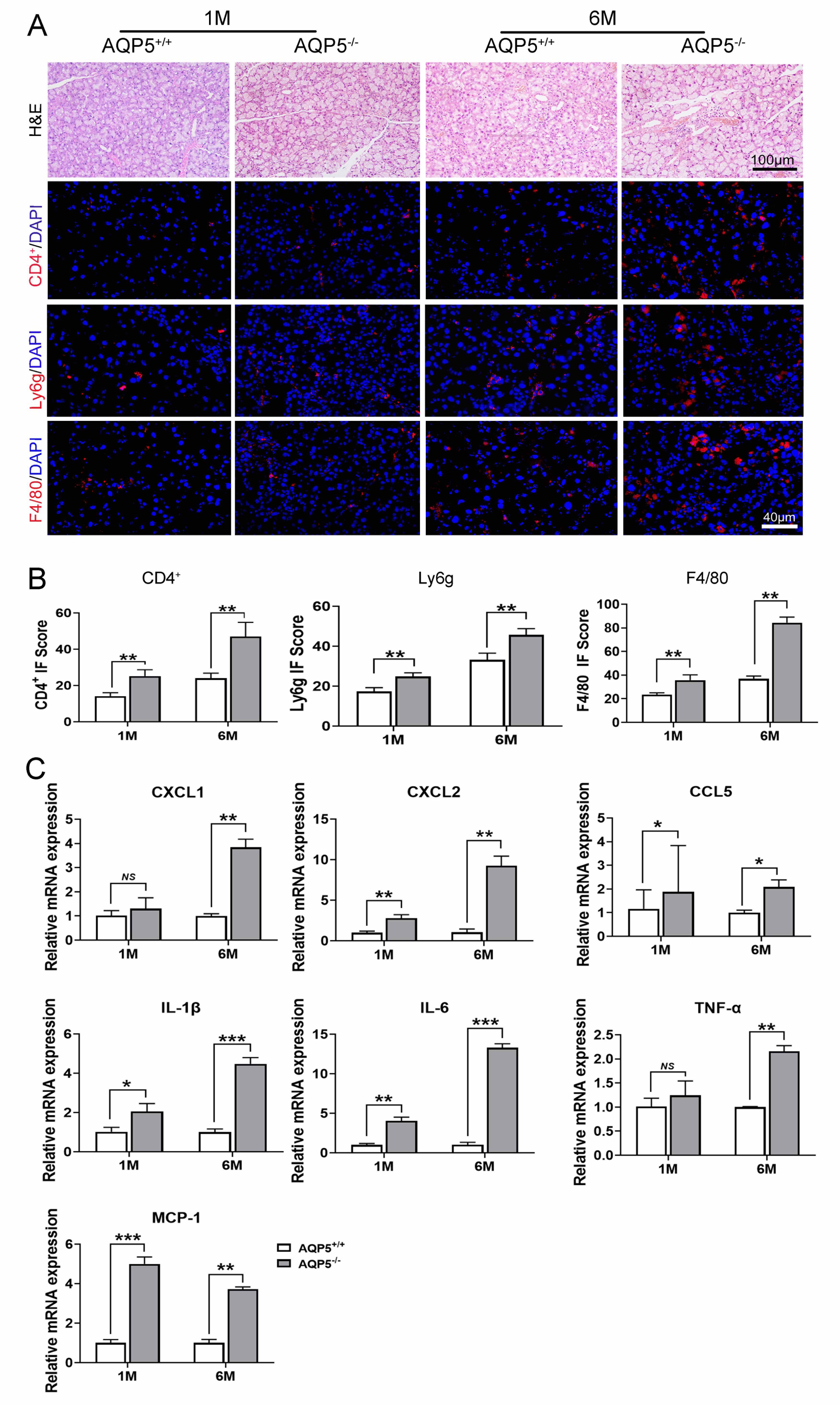
 Figure 4 of
Hu, Mol Vis 2021; 27:679-690.
Figure 4 of
Hu, Mol Vis 2021; 27:679-690.