Figure 4. Immunohistochemistry of Collagen I (Col I) displays increased collagen aggregates (yellow triangles) and an increased Col
I expression at the basement membrane (white arrows) in the Western diet (WD) fed Ossabaw mini pigs (B) compared to the Standard diet (SD) fed pigs (A). Panels C and D are negative controls where primary and secondary antibodies were excluded during antibody staining procedure.
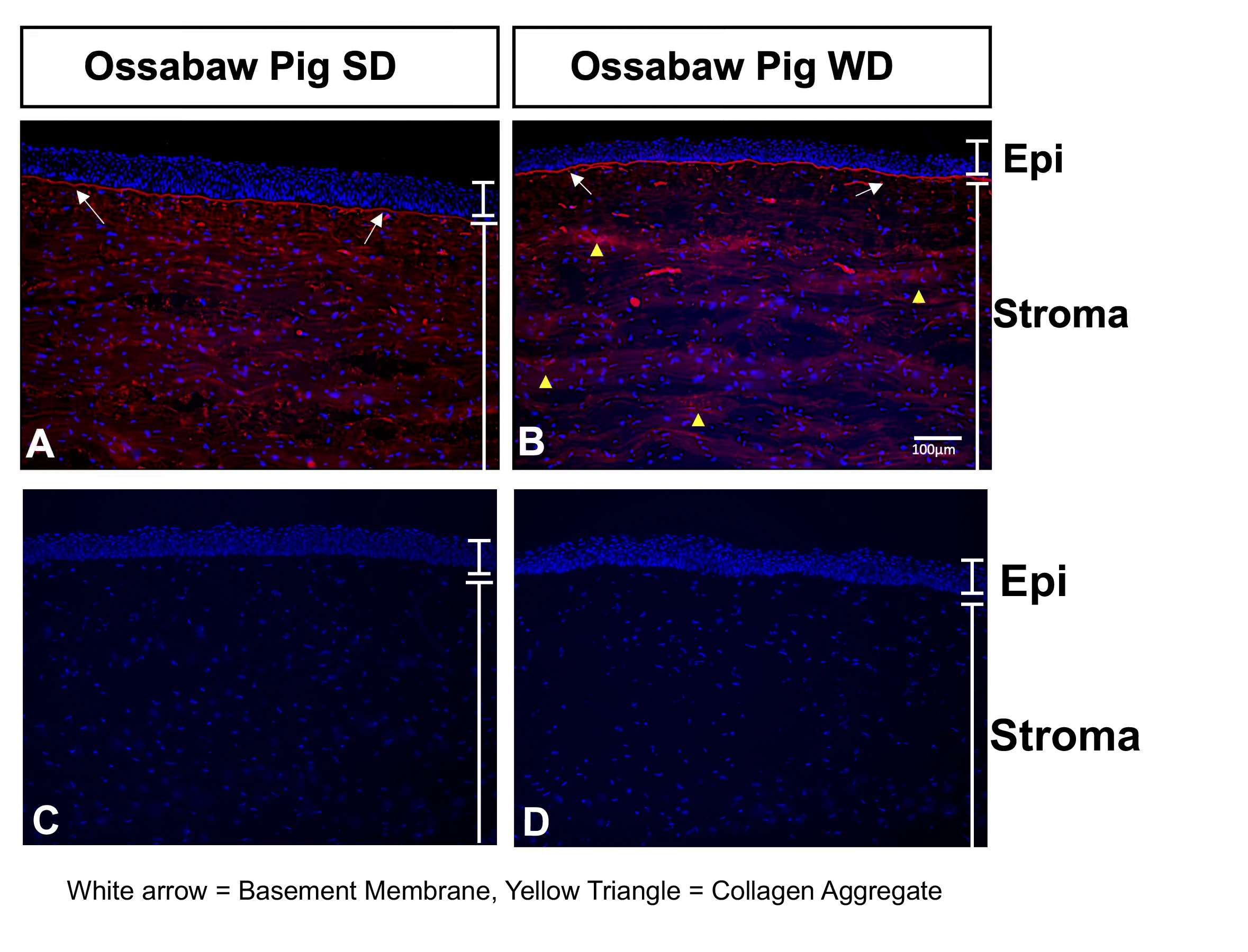
 Figure 4 of
Sinha, Mol Vis 2021; 27:666-678.
Figure 4 of
Sinha, Mol Vis 2021; 27:666-678.