Figure 3. Mason trichome staining depicts cell nuclei (black), cytoplasm (red), and collagens (blue). The Western diet (WD) fed Ossabaw
mini pigs (B) show decreased cytoplasm staining and a less even blue stain than the Standard diet (SD) fed pigs (A).
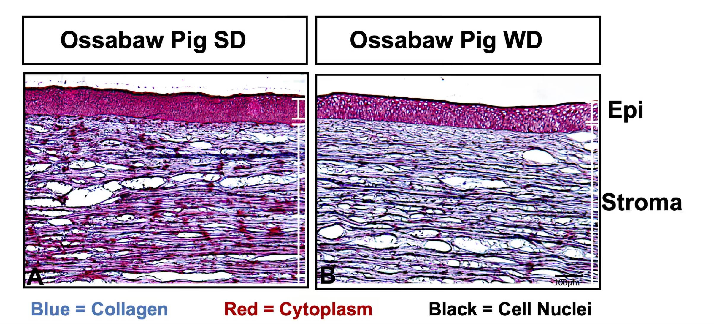
 Figure 3 of
Sinha, Mol Vis 2021; 27:666-678.
Figure 3 of
Sinha, Mol Vis 2021; 27:666-678.