Figure 3. Ocular manifestations of five FEVR patients with CNVs. OD: right eye; OS: left eye. A. Dragged disc in bilateral eyes (patient
No. 1); B. retinal fold in the right eye and dragged disc in the left eye (patient No. 2); C. deformed pupil and falciform
retinal fold in the right eye, and pupil occlusion of the left eye (patient No. 3); D. bilateral retinal fold (patient No.
4); E. corneal opacity and shallow anterior chamber in the right eye, peripheral nonperfusion area and neovascularization
of the retina in the left eye (patient No. 5).
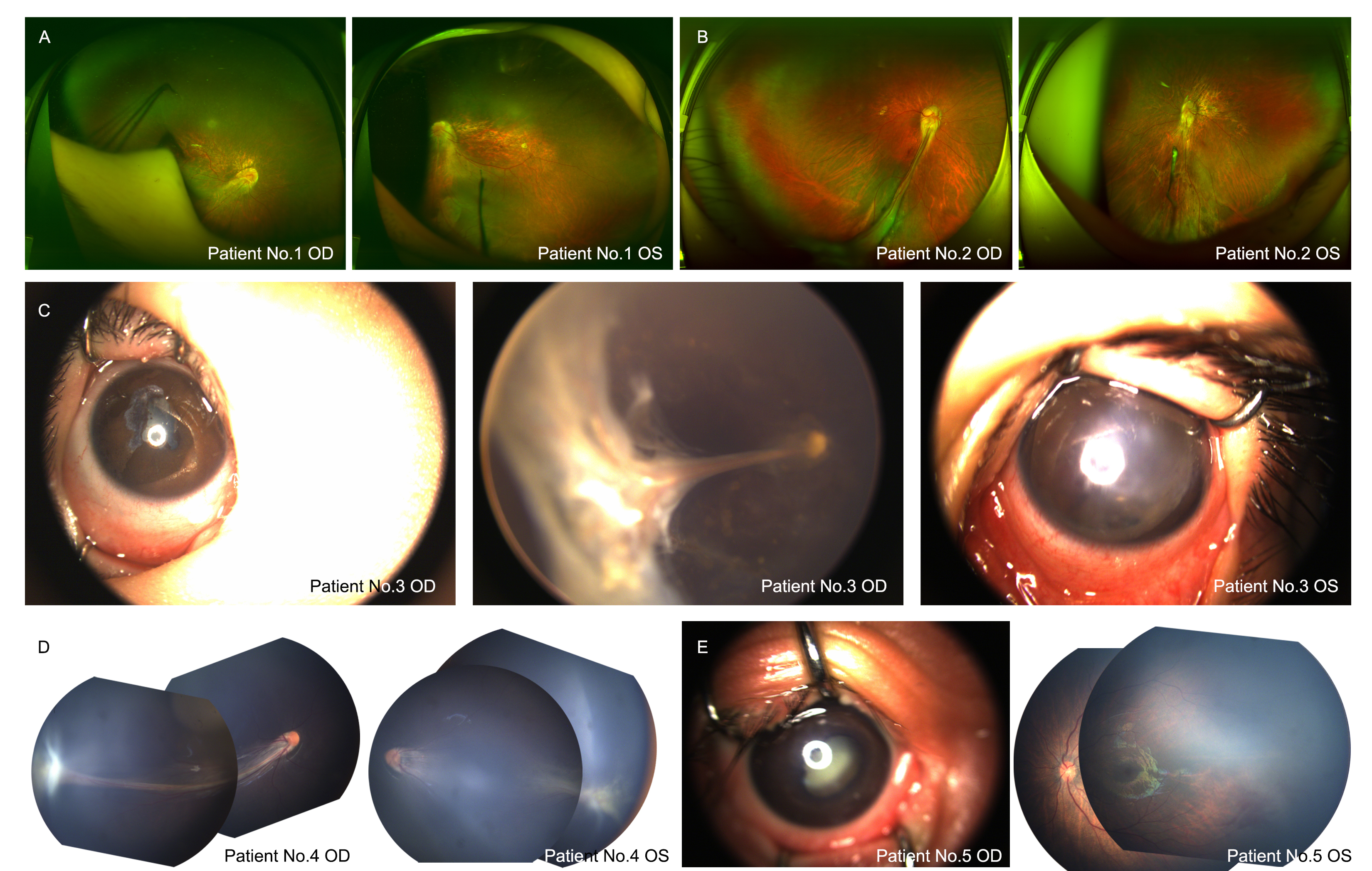
 Figure 3 of
Luo, Mol Vis 2021; 27:632-642.
Figure 3 of
Luo, Mol Vis 2021; 27:632-642.