Figure 8. Effects of ATX and LPA on the TEER and molecules associated with cell–cell contact in SCE cells. A: The TEER measured in SCE cells exposed to ATX with or without the ATX inhibitor or ROCK inhibitor at 0.5, 1, 3, 6, and 24
h after treatment. Autotaxin (ATX) statistically significantly increased the transendothelial electrical resistance (TEER)
at 3 and 6 h, and these effects were statistically significantly attenuated by the ATX inhibitor or ROCK inhibitor. B: The TEER measured in Schlemm’s canal endothelial (SCE) cells exposed to lysophosphatidic acid (LPA) with or without the
LPAR antagonist or the ROCK inhibitor at 0.5, 1, 3, 6, and 24 h after treatment. LPA statistically significantly increased
the TEER at 0.5, 1, and 3 h, and these effects were statistically significantly attenuated by the LPAR antagonist or the ROCK
inhibitor. Values are the mean ± standard deviation. (n = 4–9) #p<0.05, ##p<0.01 normal versus ATX or LPA treatment; *p<0.05, **p<0.01 ATX or LPA treatment versus each treatment. C: Immunocytochemistry in SCE cells 3 h after treatment with ATX or LPA. The left panels show staining for F-actin (red) and
β-catenin (green) merged with 4’,6-diamidino-2-phenylindole (DAPI; blue). The right panels show staining for ZO-1 (red) merged
with DAPI (blue). ATX and LPA induced rearrangement of F-actin, and upregulation of β-catenin and ZO-1 after treatment. These
effects were statistically significantly attenuated by the ATX inhibitor, the LPAR antagonist, and the ROCK inhibitor. Bar,
200 µm.
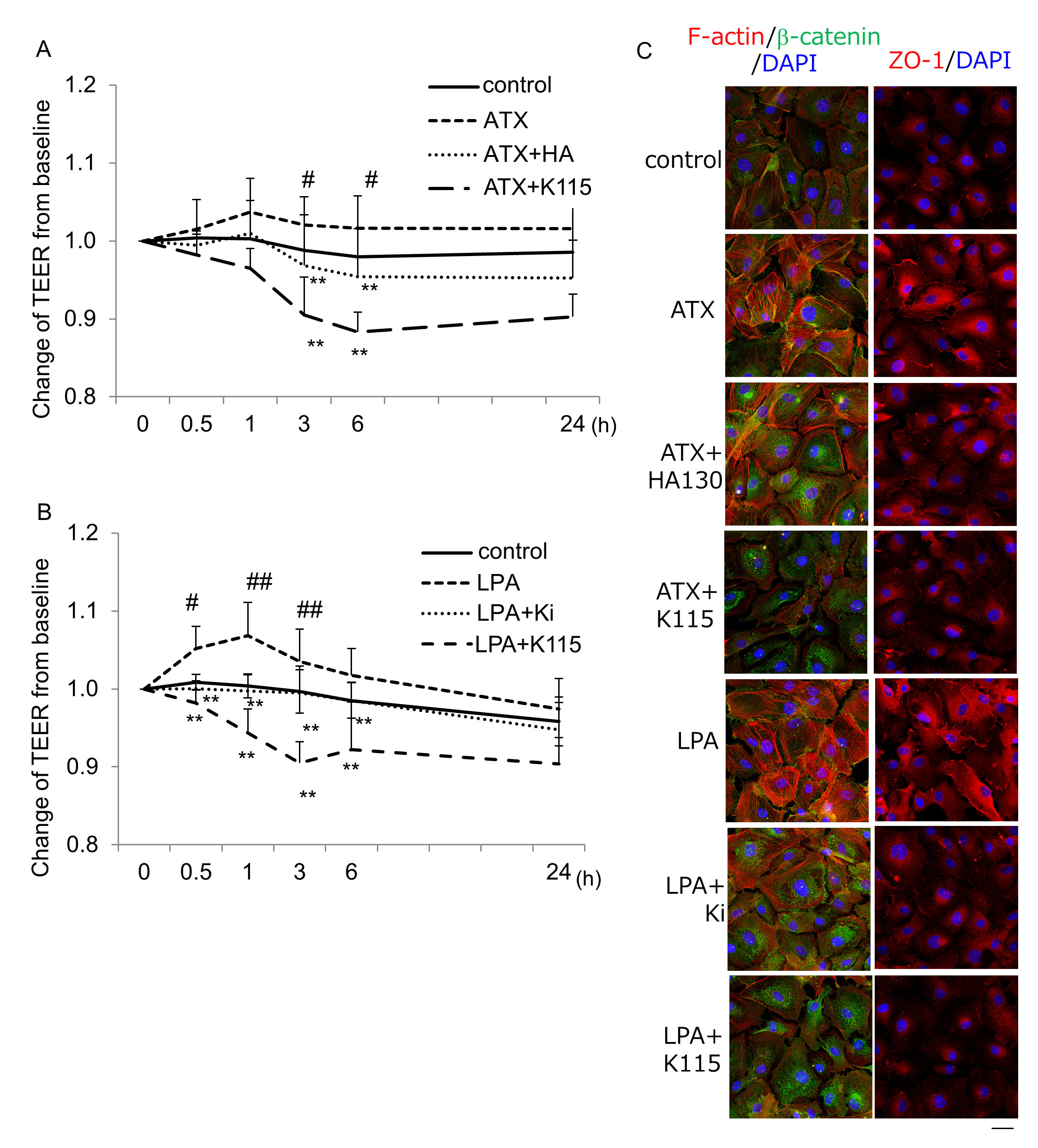
 Figure 8 of
Nakamura, Mol Vis 2021; 27:61-77.
Figure 8 of
Nakamura, Mol Vis 2021; 27:61-77.