Figure 6. Immunocytochemistry for F-actin, α-SMA, COL1A1, and fibronectin in hTM cells treated with ATX and LPA. A: Immunostaining for α-SMA (green) and COL1A1 (red) merged with 4’,6-diamidino-2-phenylindole (DAPI; blue) at 3, 6, and 24
h after treatment with 40 μM autotaxin (ATX) or 10 μM lysophosphatidic acid (LPA). LPA induced upregulation of α-SMA or COL1A1
from 3 h after treatment, and ATX induced upregulation of α-SMA or COL1A1 from 6 h after treatment. The ATX inhibitor, the
LPAR antagonist, and the ROCK inhibitor statistically significantly attenuated these changes. B: Immunostaining for fibronectin (green) and F-actin (red) merged with DAPI (blue) at 3, 6, and 24 h after treatment with
ATX or LPA. LPA and ATX induced upregulation of fibronectin from 3 h after treatment, and the ATX inhibitor, the LPAR antagonist,
and the ROCK inhibitor attenuated these changes. Bar, 200 µm.
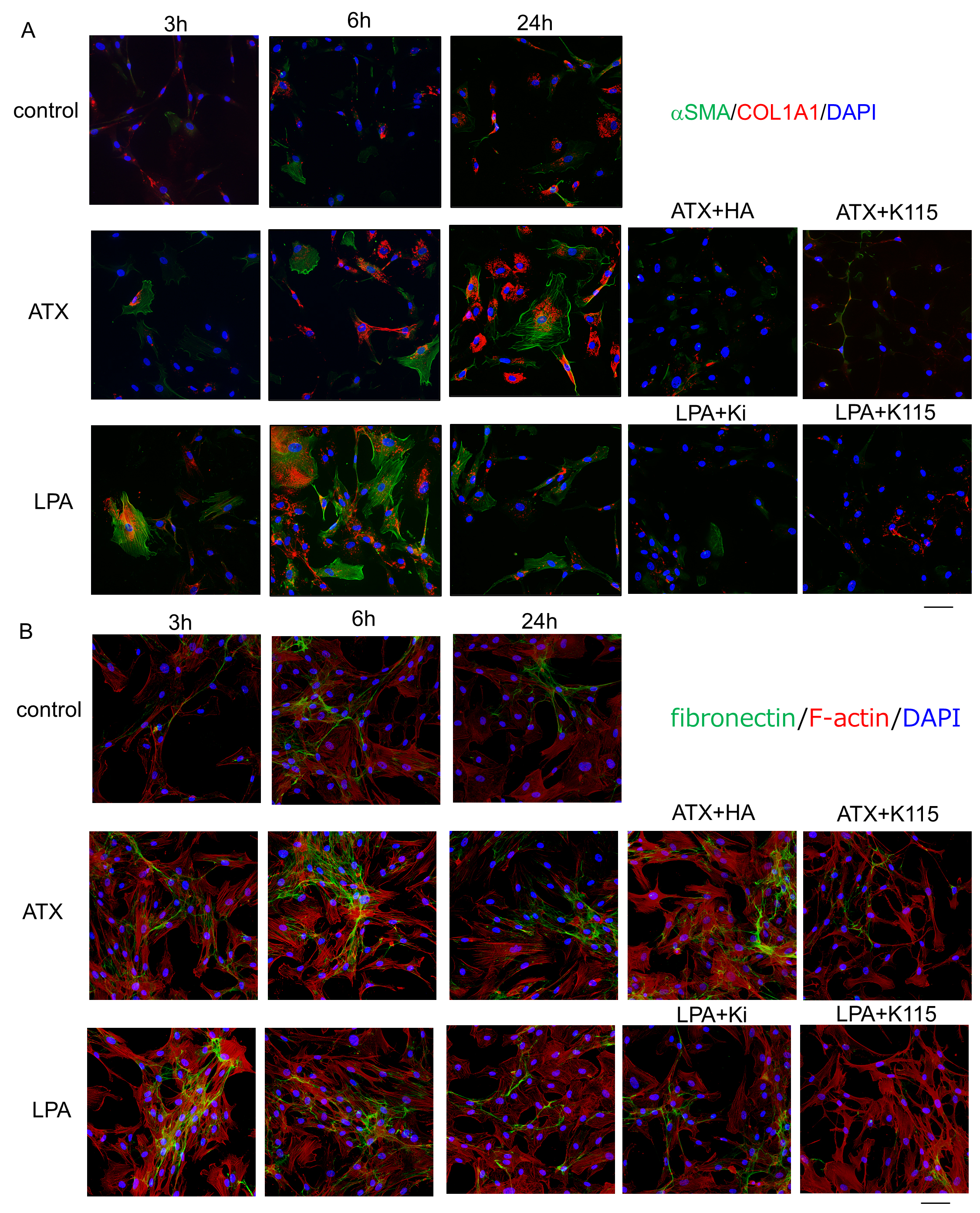
 Figure 6 of
Nakamura, Mol Vis 2021; 27:61-77.
Figure 6 of
Nakamura, Mol Vis 2021; 27:61-77.