Figure 5. Effects of TGF-β1 and TGF-β2 on monolayer permeability and TEER, and molecules associated with cell–cell contact in SCE cells.
A: Changes in Schlemm’s canal endothelial (SCE) cell monolayer permeability using 4 kDa fluorescein isothiocyanate (FITC)-dextran
are shown. SCE cells were exposed to TGF-β1 or TGF-β2 with or without the TGF-β inhibitor, and the concentrations of FITC-dextran
were measured at 1, 3, 6, and 24 h after treatment. No statistically significant differences were observed between the control
and treated cells within 24 h. B: Immunocytochemistry in SCE cells treated with 10 ng/ml TGF-β1 or TGF-β2 after 24 h. The left panels show staining for β-catenin
(green), and the right panels show staining for ZO-1 (red) merged with 4’,6-diamidino-2-phenylindole (DAPI; blue). β-catenin
and ZO-1 expression increased after treatment. Bar, 200 µm. C: The transendothelial electrical resistance (TEER) measured in SCE cells exposed to TGF-β1 or TGF-β2 with or without the
ROCK inhibitor at 1, 3, 6, and 24 h after treatment. There were no statistically significant changes in the TEER induced by
TGF-β1 or TGF-β2 compared to the control within 24 h, although the ROCK inhibitor statistically significantly decreased the
TEER at 1, 3, and 6 h. Values are the mean ± standard deviation. *p<0.05, **p<0.01, ***p<0.001, †p<0.0001.
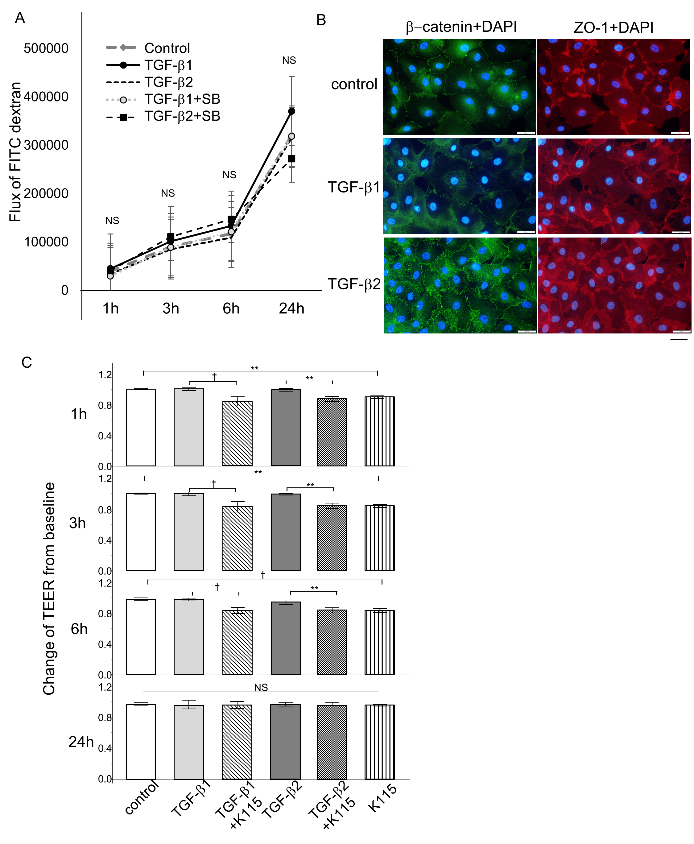
 Figure 5 of
Nakamura, Mol Vis 2021; 27:61-77.
Figure 5 of
Nakamura, Mol Vis 2021; 27:61-77.