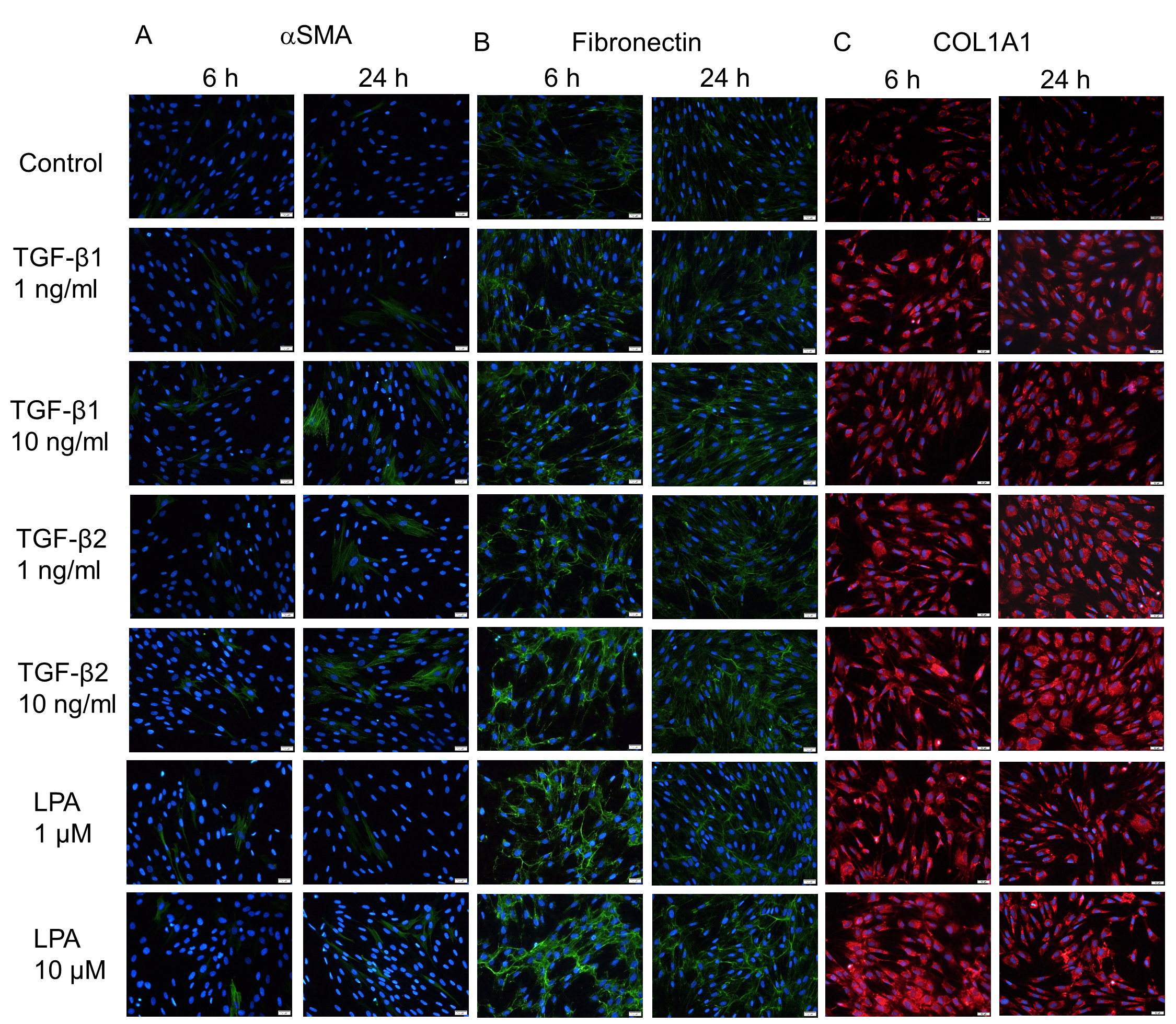
Figure 4. Immunocytochemistry of α-SMA, COL1A1, and fibronectin in hTM cells treated with TGF-β1, TGF-β2, and LPA. The human trabecular
meshwork (hTM) cells were treated with 10 ng/ml of TGF-β1 and 1 ng/ml TGF-β2 or 1 or 10 μM lysophosphatidic acid (LPA) for
6 and 24 h. The panels show cells stained for α-SMA (green, A), fibronectin (green, B), and COL1A1 (red, C) merged with 4’,6-diamidino-2-phenylindole (DAPI; blue). Bar, 200 µm.

 Figure 4 of
Nakamura, Mol Vis 2021; 27:61-77.
  Figure 4 of
Nakamura, Mol Vis 2021; 27:61-77.