Figure 1. Characterization of hTM cells. A: The image of human trabecular meshwork (hTM) cells using a contrast-phase microscope. B: The statistically significantly increased mRNA expression of myocilin was confirmed with 500 nM dexamethasone (Dex) treatment
(7 days). *p<0.05. C, D: Upregulation of myocilin was also confirmed with western blotting in hTM cells stimulated with 100 nM Dex. C: The representative bands for western blotting. D: The relative expression of myocilin to the loading control of β-tubulin (n = 3). **p<0.01. E: The panels show cells stained for 4’,6-diamidino-2-phenylindole (DAPI; blue), endothelial cell markers, and mesenchymal
markers, merged images from left to right. The hTM cells used in the study were positive for AQP-1, TIMP-3, and vimentin,
but were negative for desmin. Bar, 200 µm.
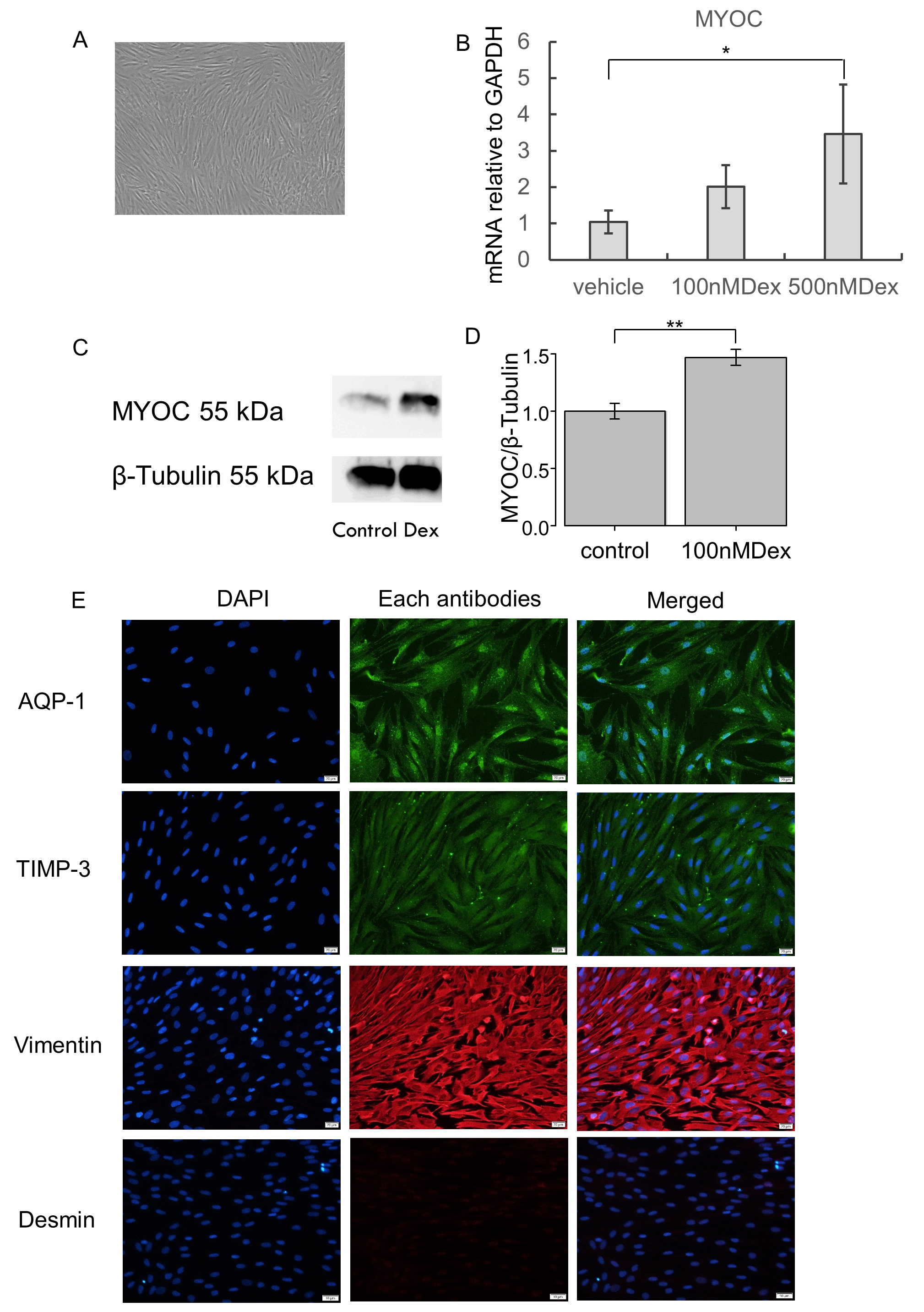
 Figure 1 of
Nakamura, Mol Vis 2021; 27:61-77.
Figure 1 of
Nakamura, Mol Vis 2021; 27:61-77.