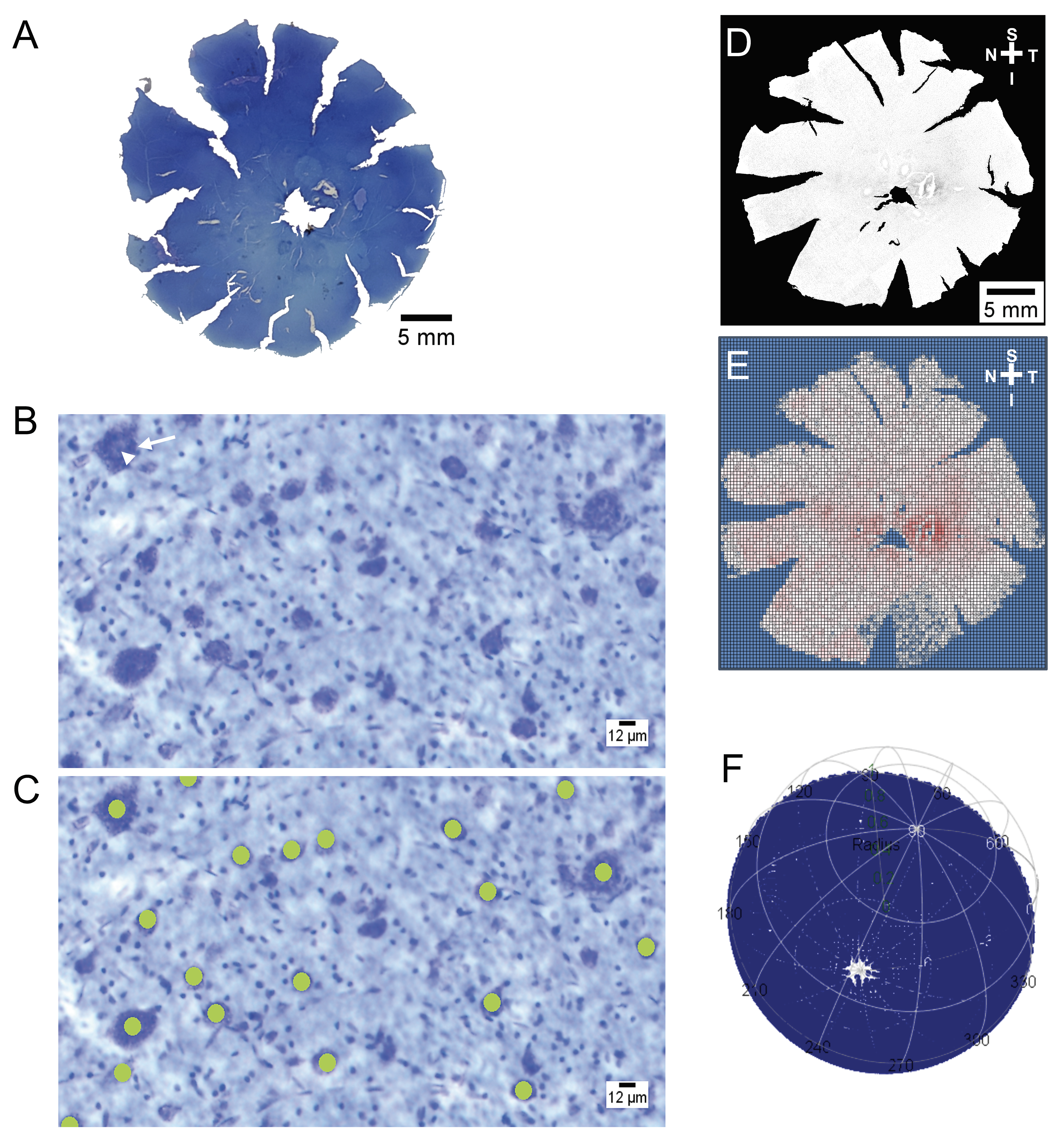
Figure 1. Wholemount preparation and imaging of feline retina. A: Image of Nissl-stained retinal wholemount with multiple peripheral retinal relief cuts made to allow the retina to lie flat.
B: Photomicrograph to illustrate the characteristic Nissl substance (white arrow), ring of cytoplasm, and nucleolus (white arrowhead)
of a properly stained retinal ganglion cell (RGC) soma. C: Photomicrographs of RGC somas illustrating the manual marking method, with 12 µm circles placed over somas that had the characteristics
outlined in (B). D: Binary image of retinal wholemount (normal wild-type [wt] retina N2) after RGC somas have been identified. “S,” “T,” “I,”
and “N” represent the directions of the superior, temporal, inferior, and nasal aspects of the retina, respectively. E: Binning of RGC soma counts within 10,000 square bins across the retina (N2; Excel, Microsoft). F: Plotting of bin locations on a three-dimensional globe (Retina, R, R Development Core Team).

 Figure 1 of
Adelman, Mol Vis 2021; 27:608-621.
  Figure 1 of
Adelman, Mol Vis 2021; 27:608-621.