Figure 2. Schematic representation of mutations detected in the PAX6 locus and FOXC1 and PITX2 genes of 20 probands. Distribution mutations were described in exons and splice sites of three genes. The frameshift (square),
missense (circle), splice-site (triangle), and nonsense (diamond) mutations are indicated. A total of one partial and one
whole gene deletions of the PAX6 gene are presented as the shaded bars. The numbers in the gray boxes refer to the exons. The functional domains PD, LR, and
HD and PST-D of the proteins are shown with the green line.
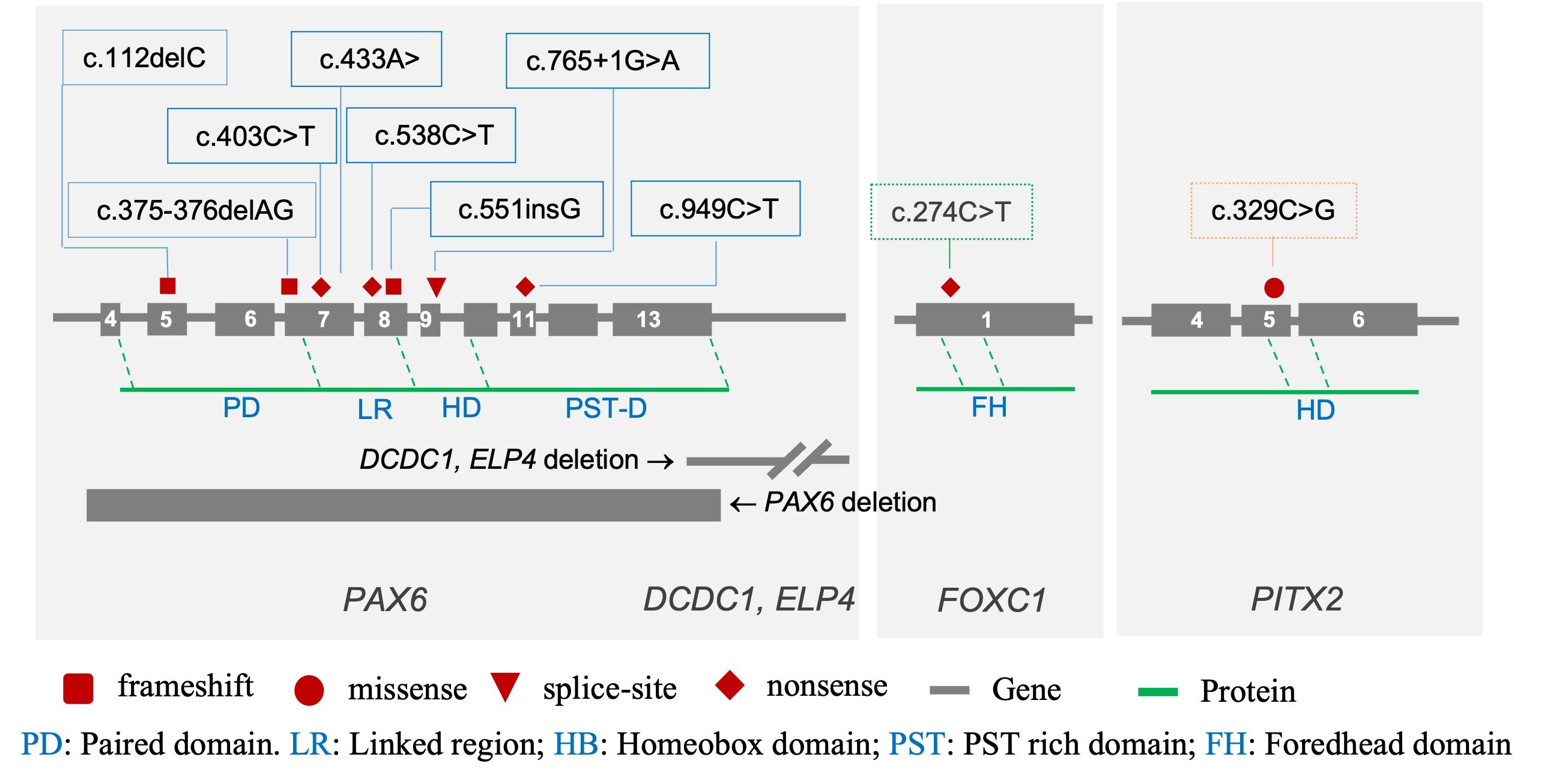
 Figure 2 of
Nguyen, Mol Vis 2021; 27:555-563.
Figure 2 of
Nguyen, Mol Vis 2021; 27:555-563.