Figure 4. Patient P5. A, B, C, D, E: Color fundus photograph and FA of the right eye of patient P5. The color fundus photograph shows a well-positioned scleral
buckle and three peripheral retinal capillary hemangioblastoma (RCHs) (A, B, C) with scarring on the surrounding retina due to the cryotherapy (2 weeks after surgery, in 2010). On the right, the fluorescein
angiography (FA) images highlight the blood supply of the three RCHs after cryotherapy and laser. A: Peripheral RCH located at 11 o’clock. B: Peripheral RCH at 8 o’clock. C: Peripheral RCH at 6 o’clock. D: In 2019, ultrawide field (UWF) color fundus photography shows the effect of the scleral buckle and the peripheral three
lesions at 6, 8, and 11 o’clock. E: Color fundus photograph and FA of the right eye show a juxtapapillary fibrovascular membrane (white arrowheads).
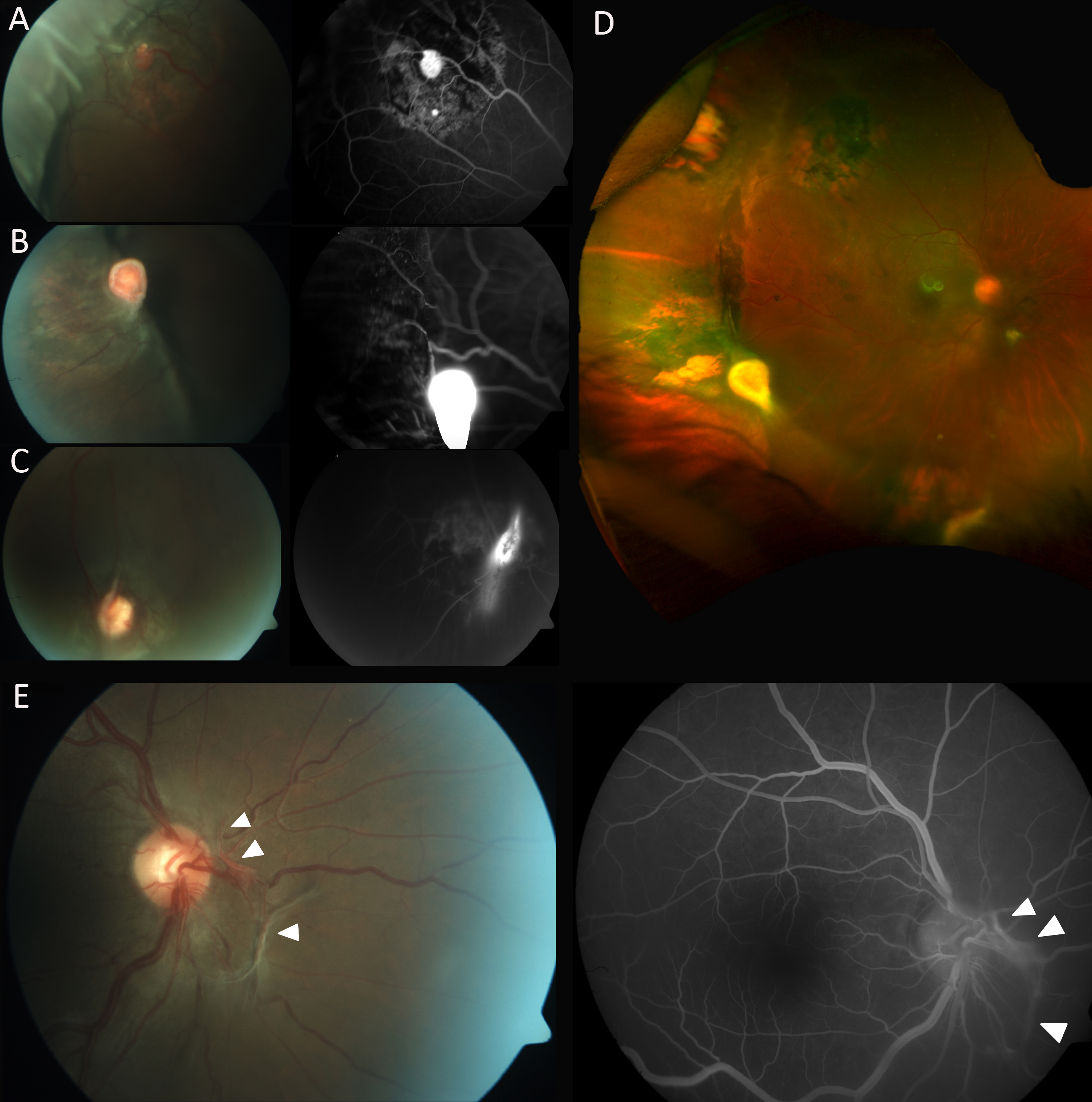
 Figure 4 of
Murro, Mol Vis 2021; 27:542-554.
Figure 4 of
Murro, Mol Vis 2021; 27:542-554.