Figure 3. Patient P2. FA of the right eye of patient P2 shows the RCH (hyperfluorescence round lesion) before (B) and after (D) multiple laser sessions. The color fundus photograph shows the retinal capillary hemangioblastoma (RCH) after the first
laser session (C). The optical coherence tomography (OCT) scan shows the irregularities of the inner retinal surface before treatment (pseudohole;
2009; A). Two months after the beginning of the laser treatment, the OCT scan shows a lamellar macular hole (E) and then a full-thickness macular hole (FTMH) in 2010 (F). In particular, hyperreflective abnormalities above the RPE can be seen in the bed of the hole (F) which appear as round yellowish lesions in the color fundus photograph (G). The yellow lesions are particularly evident in the magnified foveal image performed in 2012 which shows the increase in
the number and size of the yellowish dots during follow-up (G). The OCT scan (H) shows the closure of the FTMH after vitrectomy. At the last follow-up visit in 2019, the ultrawide field (UWF) color fundus
photograph (I) shows the RCH regression.
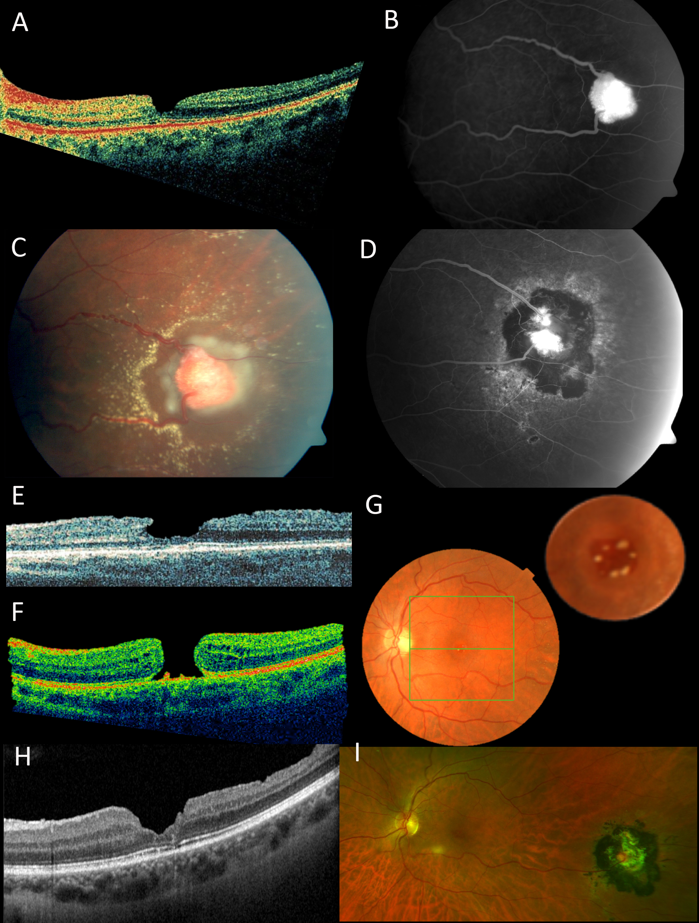
 Figure 3 of
Murro, Mol Vis 2021; 27:542-554.
Figure 3 of
Murro, Mol Vis 2021; 27:542-554.