Figure 2. Patient P1. Color fundus photograph (A) shows the single peripheral RCH after one session of laser photocoagulation treatment (2015). Fluorescein angiography (FA;
B) displays the feeder vessels and the early hyperfluorescence of the retinal capillary hemangioblastoma (RCH) lesion (2015).
The ultrawide field (UWF) color fundus photograph (C) shows the inferonasal location of the RCH (2019). During follow-up, the lesion remained stable. UWF autofluorescence imaging
(D) shows the retinal scar following the treatment (hypoautofluorescence lesion).
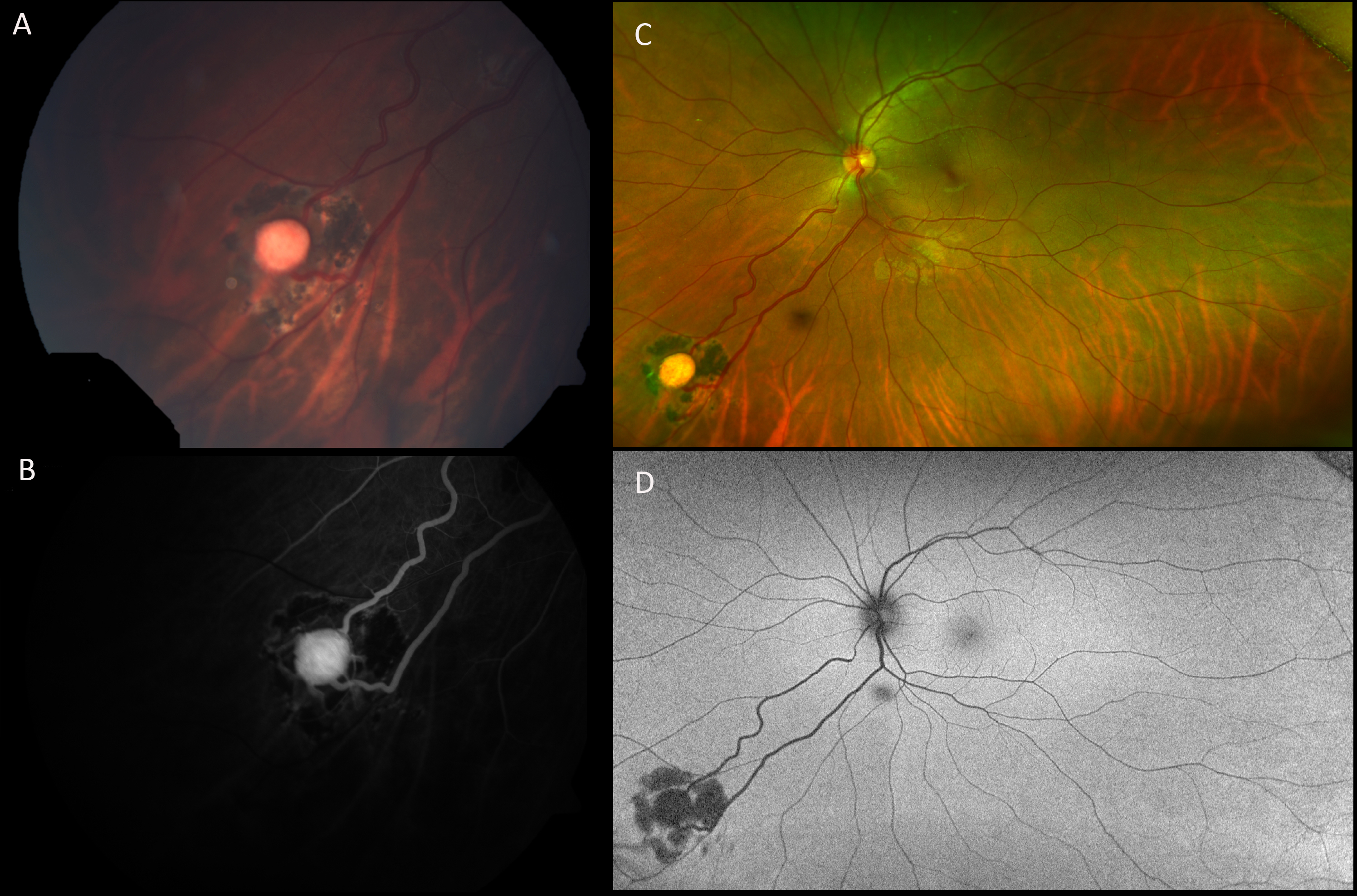
 Figure 2 of
Murro, Mol Vis 2021; 27:542-554.
Figure 2 of
Murro, Mol Vis 2021; 27:542-554.