Figure 5. Fluorescein angiography images of probands’ parents who carry the KIF11 mutation p. H718L. Due to the normal posterior retinal vessels, their visual acuity was completely normal. However, the avascular
area (red asterisk) and peripheral aberrant vascularization (yellow arrow) were detected after the fluorescein angiography
examination. OD and OS represent the right and left eye, respectively.
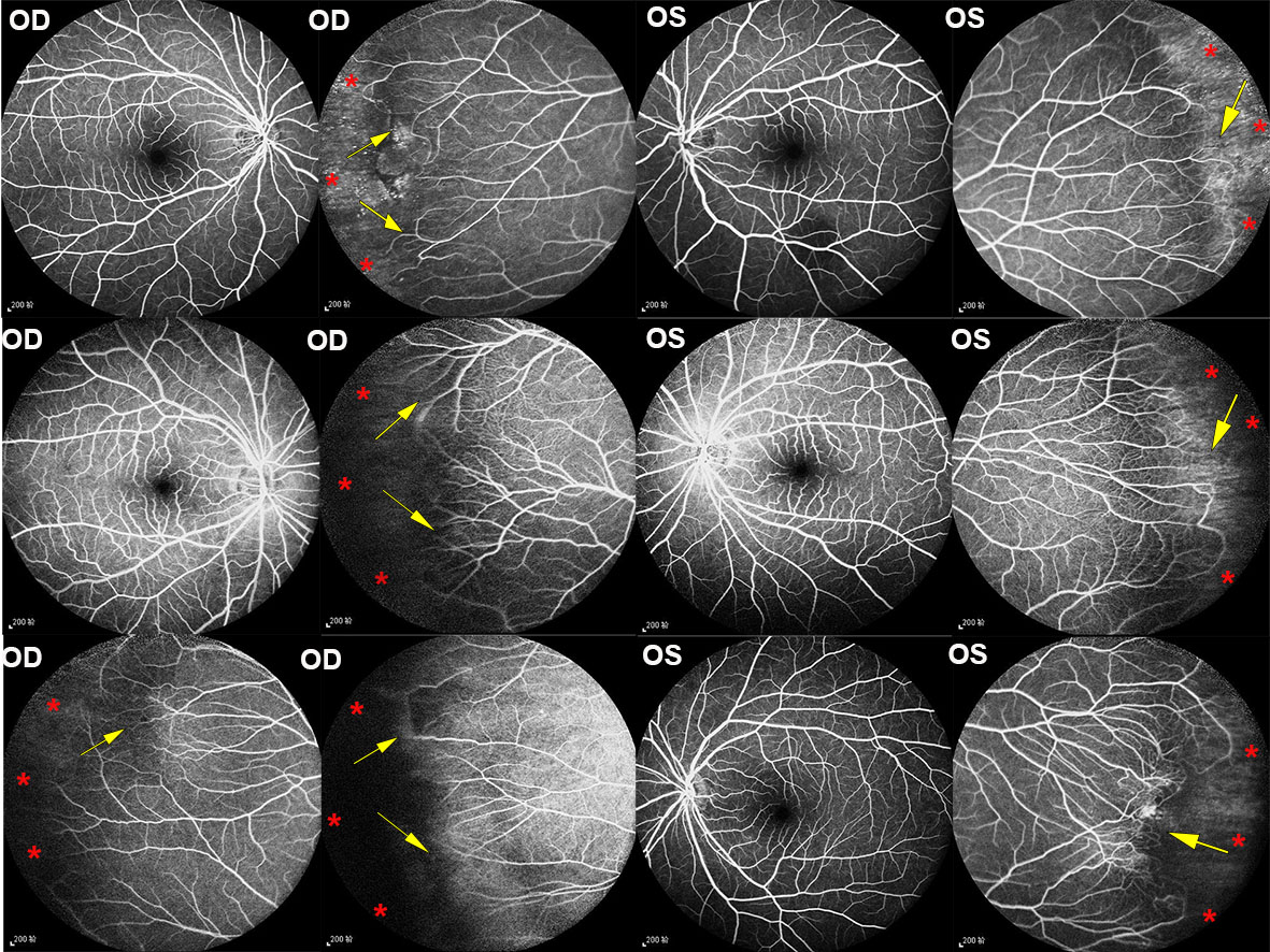
 Figure 5 of
Wang, Mol Vis 2021; 27:528-541.
Figure 5 of
Wang, Mol Vis 2021; 27:528-541.