Figure 4. Fundus images of proband 12 (LRP5 mutation p. R494W, LRP5 mutation p. R41W, and KIF11 mutation p. H718L). A: Avascular zone and exudation are observed in the right eye. B: The left eye exhibits total retinal detachment with a retrolenticular fibrotic mass. C, D: During follow-up, falciform retinal detachment developed in the right eye, and the left eye remained total retinal detachment.
OD and OS represent the right and left eye, respectively. E, F: Peripheral aberrant vascularization and avascular zone are observed in the proband’s affected father (LRP5 mutation p. R41W, KIF11 mutation p. H718L). G, H: The affected mother presents as avascular zone and peripheral aberrant vascularization (LRP5 mutation p. R494W).
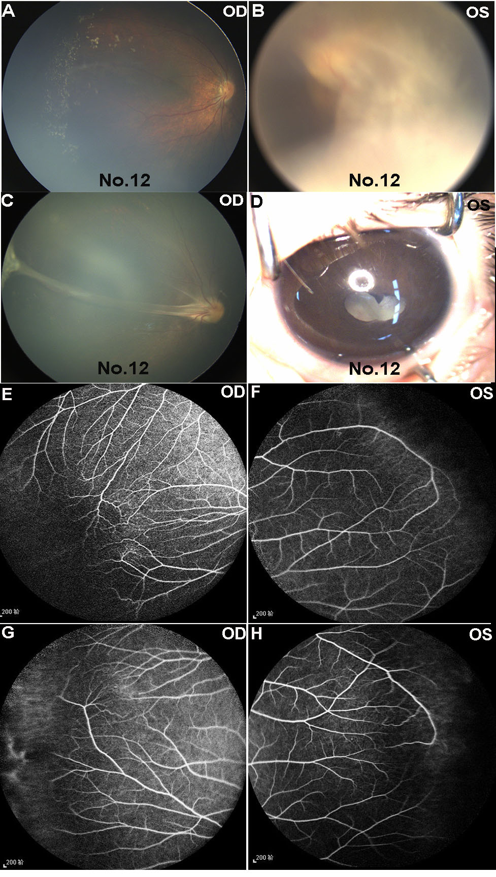
 Figure 4 of
Wang, Mol Vis 2021; 27:528-541.
Figure 4 of
Wang, Mol Vis 2021; 27:528-541.