Figure 2. Fundus photo and SD-OCT of the proband (I:2). Fundus picture of the right (A) and left (B) eyes, showing normal optic discs and symmetric macular coloboma-like excavations, consistent with North Carolina macular
dystrophy (NCMD) grade 3. Spectral domain-optical coherence tomography (SD-OCT) of the right eye (C) and of the left eye (D) illustrate a macular coloboma-like lesion with an absence of the RPE and intrachoroidal fluid representing a lacuna.
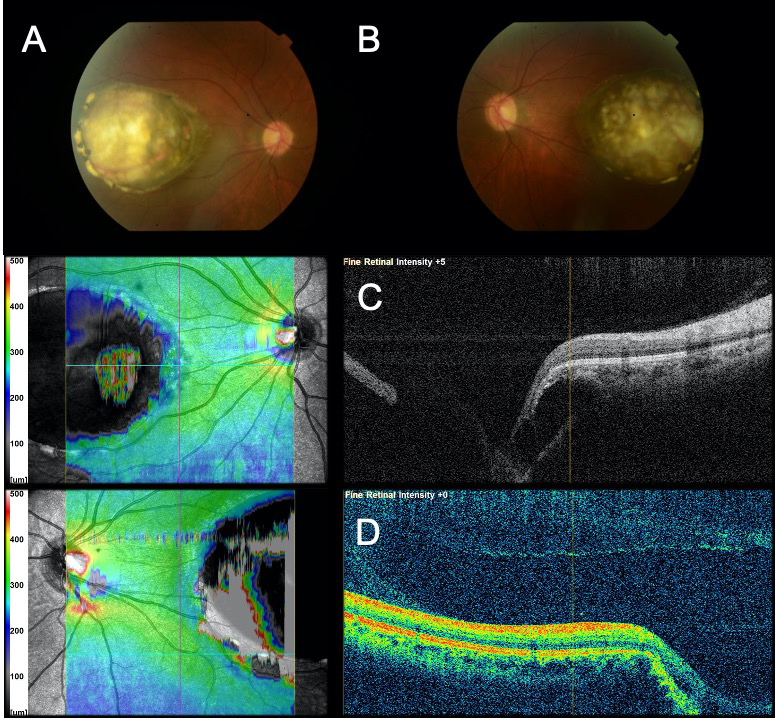
 Figure 2 of
Small, Mol Vis 2021; 27:518-527.
Figure 2 of
Small, Mol Vis 2021; 27:518-527.