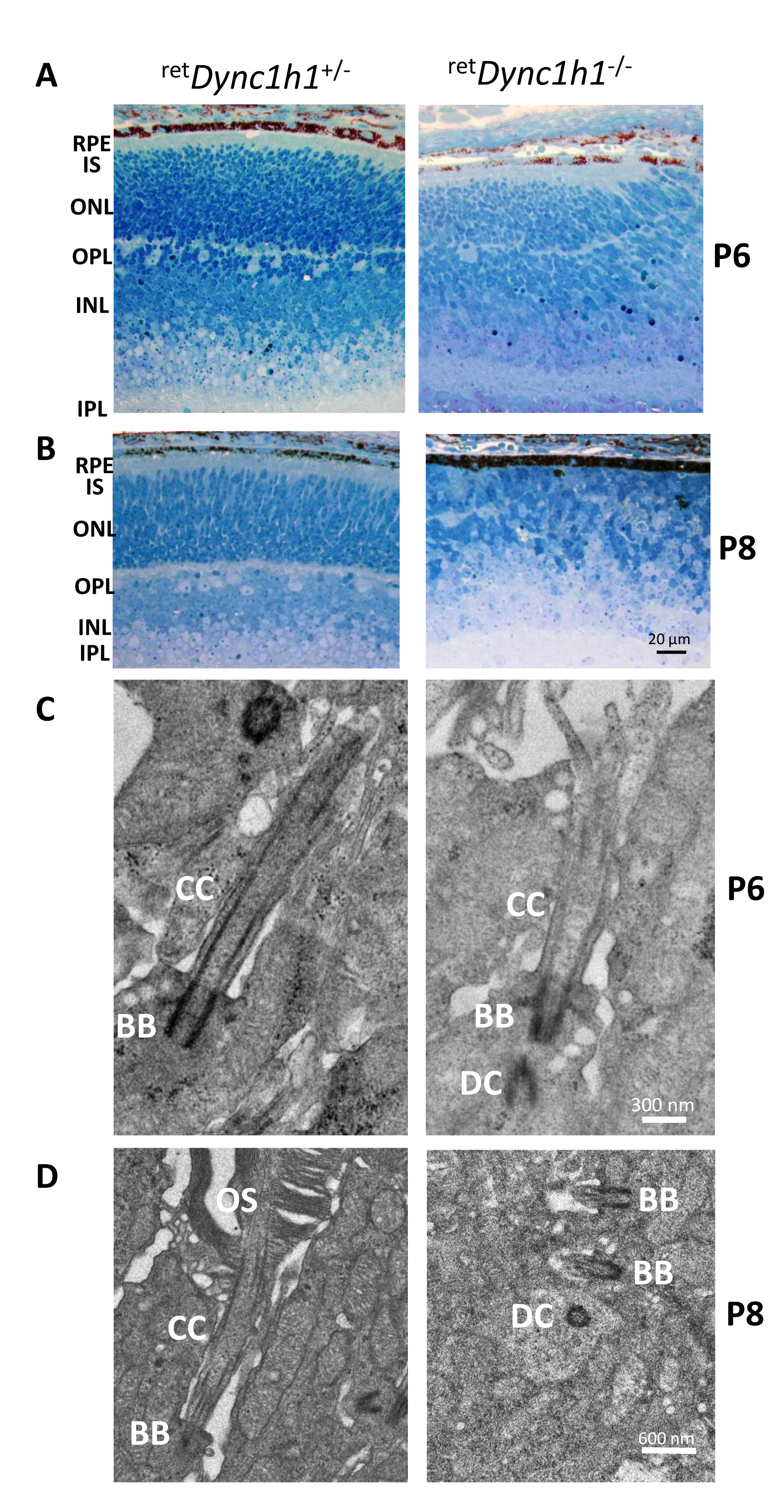
Figure 4. Defective
retDync1h1−/− retina lamination and impaired ciliogenesis.
A, B: Plastic sections of the central retina near the optic nerve of heterozygous control (left) and
retDync1h1−/− (right) mice at P6 and P8. Sections are stained with methylene blue-Azure II (Richardson’s) to demonstrate the retina layers.
RPE, retinal pigmented epithelium; IS, inner segment; ONL, outer nuclear layer; OPL, outer plexiform layer; INL, inner nuclear
layer; IPL, inner plexiform layer; GCL, ganglion cell layer. Scale bar = 20 μm.
C: Representative ultrastructure of connecting cilia emanating from heterozygous control (left panel) and
retDync1h1−/− basal bodies (right panel) at P6. Note the presence of the daughter centriole (DC), basal body (BB) docking to the membrane,
and connecting cilium (CC) elaboration in the
retDync1h1−/− photoreceptor, scale bar = 0.3 μm.
D: Axonemes and connecting cilia are absent at P8, scale bar = 0.6 μm. Left panel, heterozygous control; right panel,
Dync1h1 knockout mouse. Modified from [
72] with permission from
PLOS One.  Figure 4 of
Dahl, Mol Vis 2021; 27:506-517.
Figure 4 of
Dahl, Mol Vis 2021; 27:506-517.