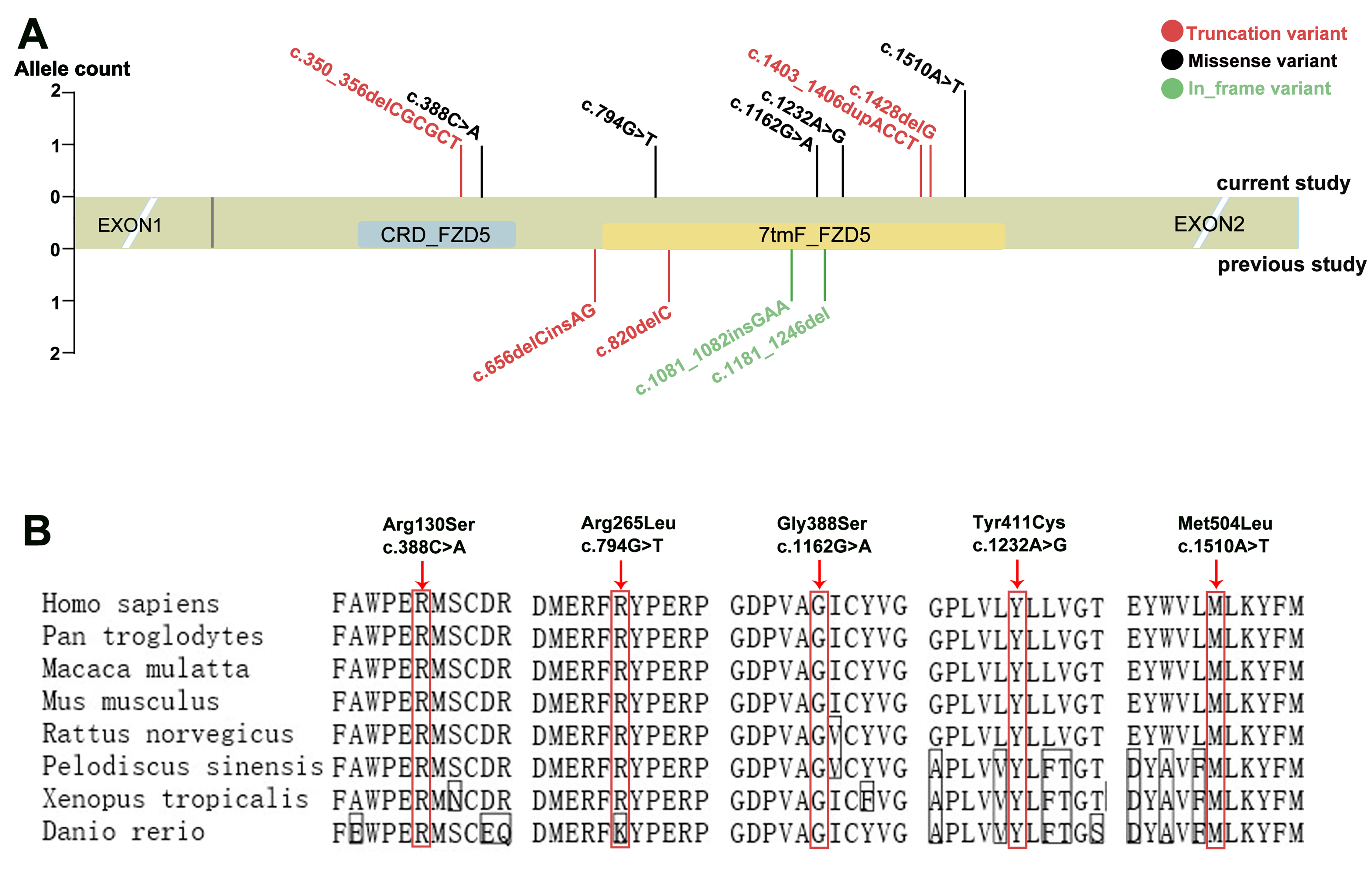
Figure 4. Distribution and conservation analysis of
FZD5 variants.
A: The allele count and distribution of all
FZD5 variants in the mRNA sequence in the current study and previous studies (Ref.
NM_ 003468.3). The light green rectangle represents the mRNA sequence of
FZD5. The potential pathogenic variants in the current study are shown above the structure of the mRNA sequence, while variants
in other previous studies are shown below the structure of the mRNA sequence. The red, black, and green variants represent
truncation variants, missense variants, and in-frame variants, respectively. The blue area represents the extracellular cysteine-rich
Wnt-binding domain, the yellow area represents seven transmembrane Frizzled domains, and the two blank areas before and after
the sequence represent the 5′UTR and 3′UTR, respectively. The blank area between two slashes indicates the partial sequences
of the 5′UTR and 3′UTR.
B: The conservation analysis of five missense variants identified in our study. Sequence alignment of the sequences of
Homo sapiens (humans) and seven other seven species, including
Pan troglodytes (chimpanzee),
Macaca mulatta (monkey),
Mus musculus (house mouse),
Rattus norvegicus (rat),
Pelodiscus sinensis (Chinese soft-shelled turtle),
Xenopus tropicalis (Western clawed frog), and
Danio rerio (zebrafish). The five missense variants were located in the conserved region of the FZD5 protein among eight species.
 Figure 4 of
Jiang, Mol Vis 2021; 27:50-60.
Figure 4 of
Jiang, Mol Vis 2021; 27:50-60.