Figure 3. Representative fundus photographs from patients with FZD5 variants. A-E: Inferior chorioretinal and optic disc hypoplasia presented in both eyes from two patients (12467-I:1 and 13706-I:1). F: The fundus photograph showed typical features of myopic fundus: tessellated retina and partial foveal atrophy in one patient
(13706-II:1). G: Inferior chorioretinal hypoplasia was observed in the lower left area of the fundus in the right eye from patient 9574-II:1.
H: Uveal coloboma and tessellated fundus, located between the coloboma and optic disc, were observed in patient 1267-II:1.
I: A normal fundus photo in individual 5485-I:2.
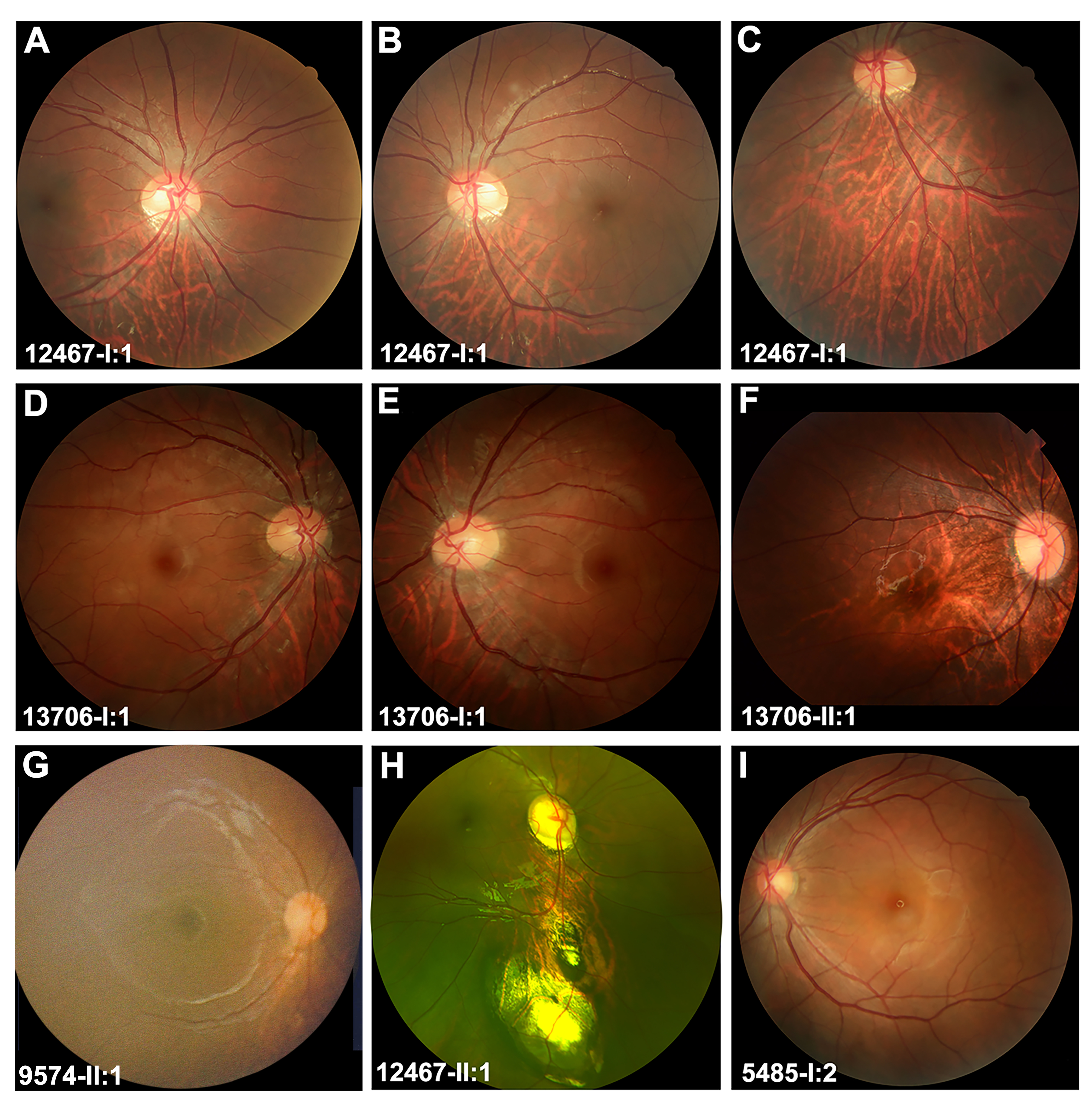
 Figure 3 of
Jiang, Mol Vis 2021; 27:50-60.
Figure 3 of
Jiang, Mol Vis 2021; 27:50-60.