Figure 3. The image processing procedure for large and small vessels. The raw optical coherence tomography angiography (OCTA) image
of the superficial vascular plexus (SVP) (A) with a field of view 6 × 6 mm2 was processed to extract the large vasculature with a diameter of about >25 µm (B), and the small vessels (C) were skeletonized for fractal analysis. For the deep vascular plexus (DVP) (D), the large vessels (E) were removed, and the remaining small vessels were skeletonized (F) for analysis.
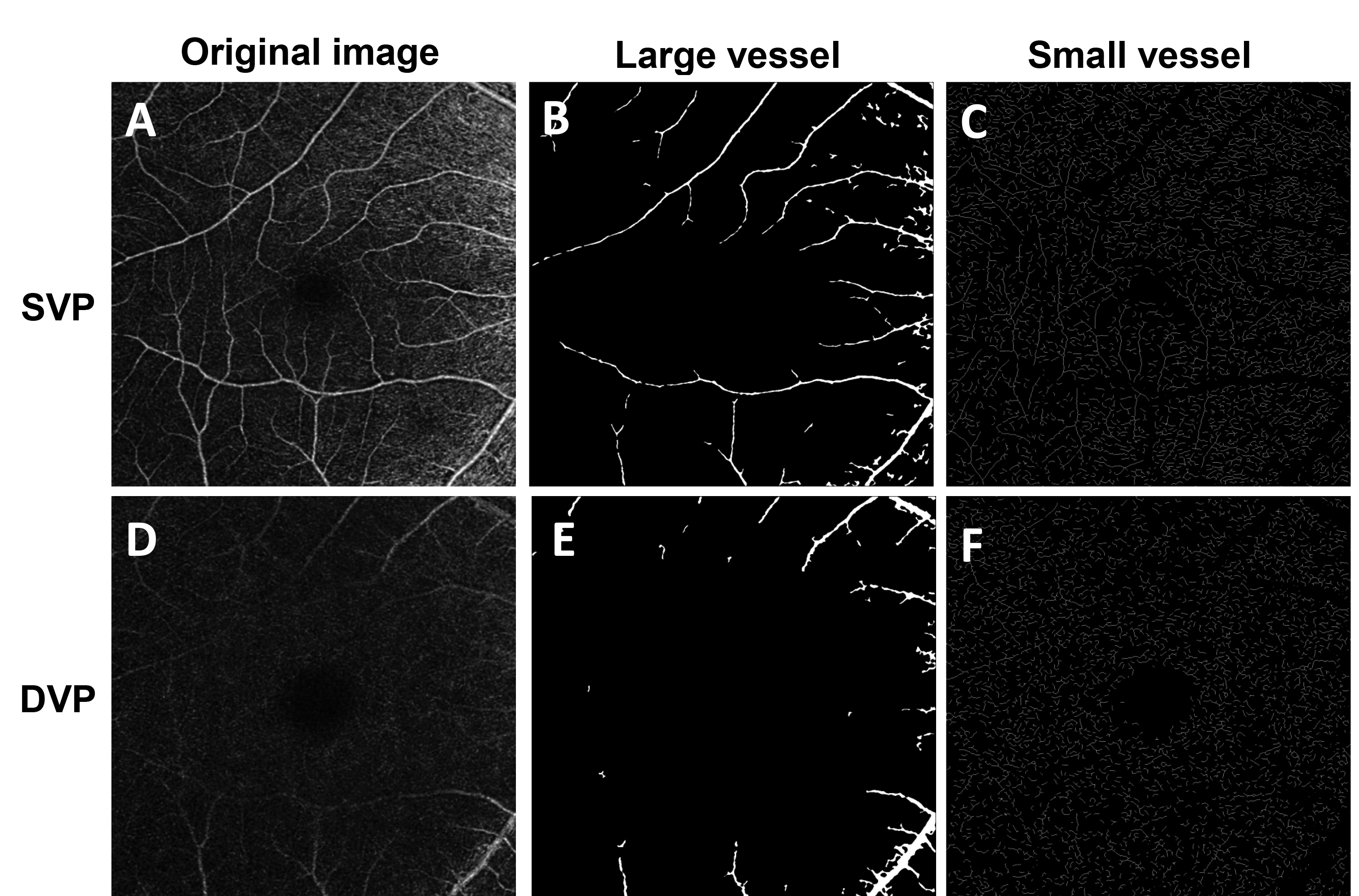
 Figure 3 of
Hu, Mol Vis 2021; 27:466-479.
Figure 3 of
Hu, Mol Vis 2021; 27:466-479.