Figure 1. Pre- and postoperative 6 × 6 mm2 OCTA scan images. A–D: The images show the retinal vascular network (RVN) preoperatively and at the three follow-up visits (1 week, 1 month, and
3 months postoperatively). E–H: The images show the superficial vascular plexus (SVP). I–L: The images show the deep vascular plexus (DVP) at the three time points described above.
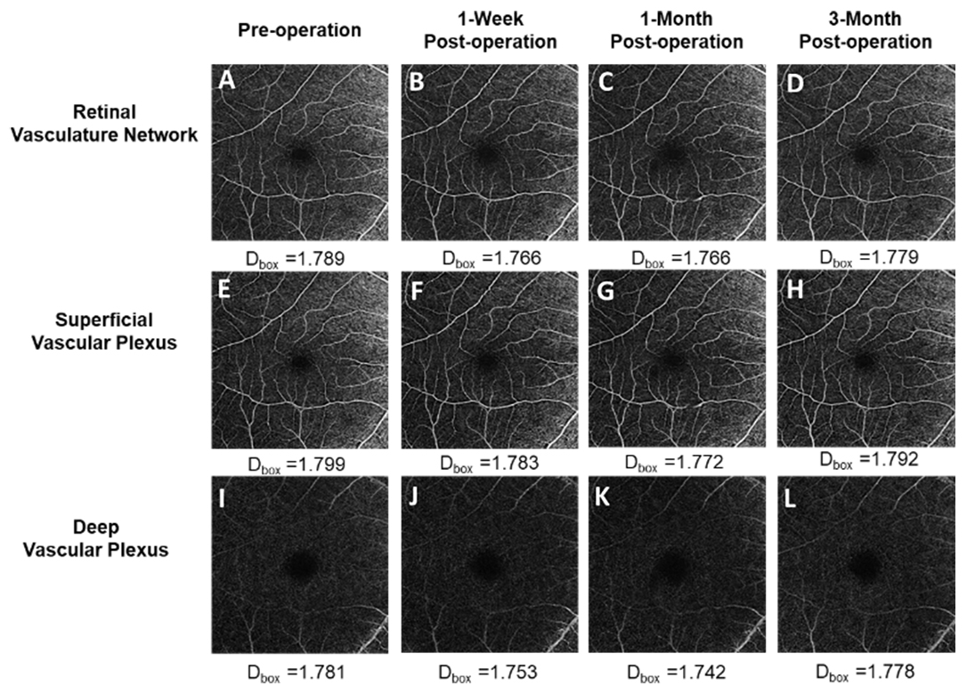
 Figure 1 of
Hu, Mol Vis 2021; 27:466-479.
Figure 1 of
Hu, Mol Vis 2021; 27:466-479.