Figure 9. Retrograde-labeled RGCs in the retina at 14 days after IR injury. The picture of the retina was taken at bitemporal 1.5 μm
from the optic nerve. A, D: Sham group; (B, E) minocycline treatment (Mino) group; (C, F) no treatment (NT) group. A–C show only cells with active retrograde transport were stained with 1,1’-dioctadecyl-3,3,3′,3′-tetramethylindocarbocyanine
perchlorate (DiI; red). D–F show the merged images with cell nuclei stained with 4',6-diamidino-2-phenylindole (DAPI; blue). The number of retinal ganglion
cells (RGCs) decreased markedly after IR injury compared with the sham group, while the minocycline prevented the numbers
of RGCs from decreasing. Scale bar: 50 μm. G: The average cell numbers of retrograde-labeled RGCs for each visual field in each group (n = 6) at 14 days after IR. Sham:
sham group without IR injury; NT-IR: NT group with IR injury; Mino-IR: Mino group with IR injury. ***p<0.001; **p<0.01.
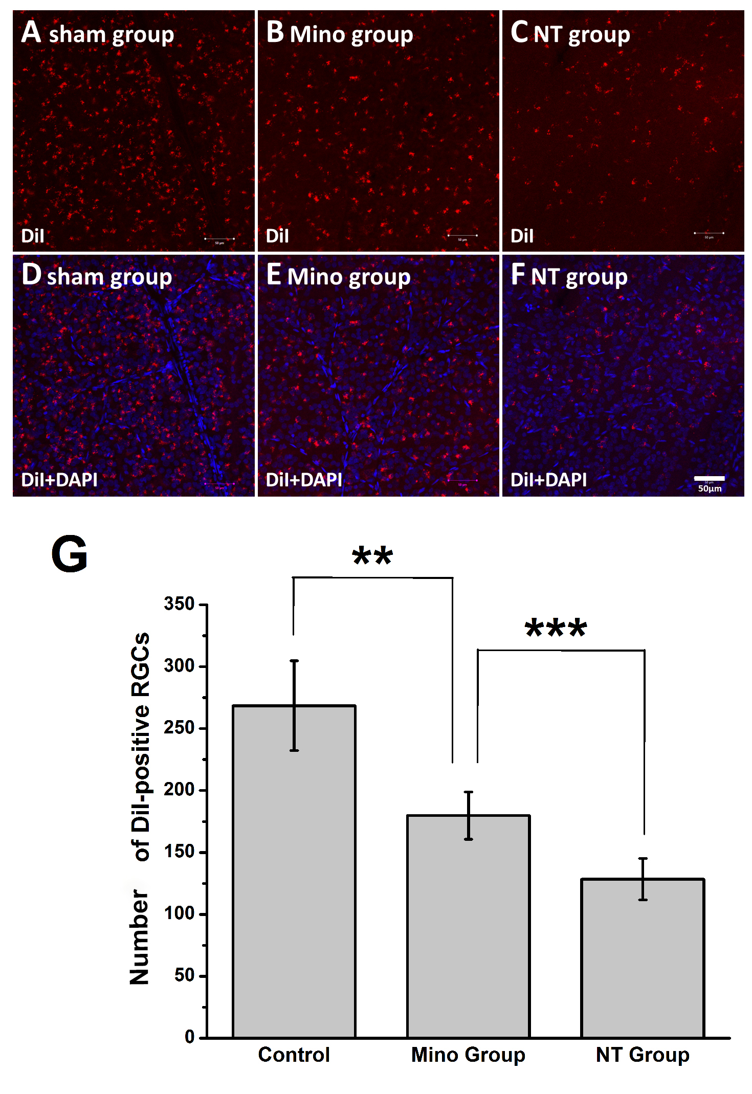
 Figure 9 of
Li, Mol Vis 2021; 27:438-456.
Figure 9 of
Li, Mol Vis 2021; 27:438-456.