Figure 6. The thickness of each retinal layer at different locations measured using H&E staining at 3 days and 14 days after IR. A, B: Retinal nerve fiber layer (RNFL) and ganglion cell layer (GCL; RNFL + GCL). C, D: Inner plexiform layer (IPL). E, F: Inner nuclear layer (INL). G, H: Outer nuclear layer (ONL). n = 6, ***p<0.001.
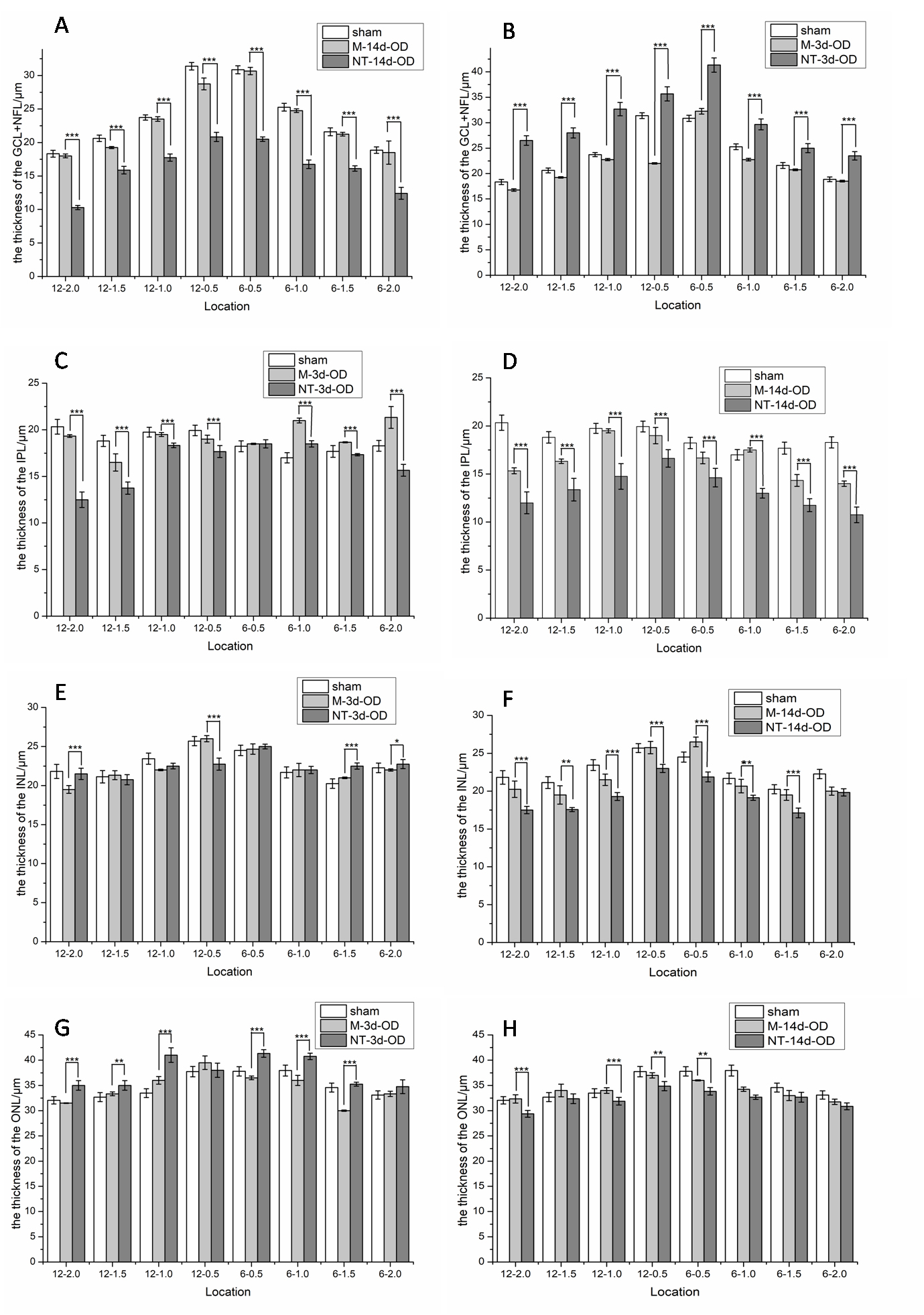
 Figure 6 of
Li, Mol Vis 2021; 27:438-456.
Figure 6 of
Li, Mol Vis 2021; 27:438-456.