Figure 2. Fundus photography and retinal vessels (stained with IB4) in the Mino group and the no treatment (NT) group. The level of
retinal vessel circuity and dilation was reduced in the minocycline treatment (Mino) group (A, D) compared to the NT group (B, E). The retinal vessels of the fellow eyes did not change after treatment (C, F). In A and B, black arrows point out the vessel’s circuity and dilation. In D, E, and F, IB4 staining shows small blood vessel morphology. (In panel E, the red arrow points to the fluorescence leakage suggesting that the blood–retinal barrier was damaged).
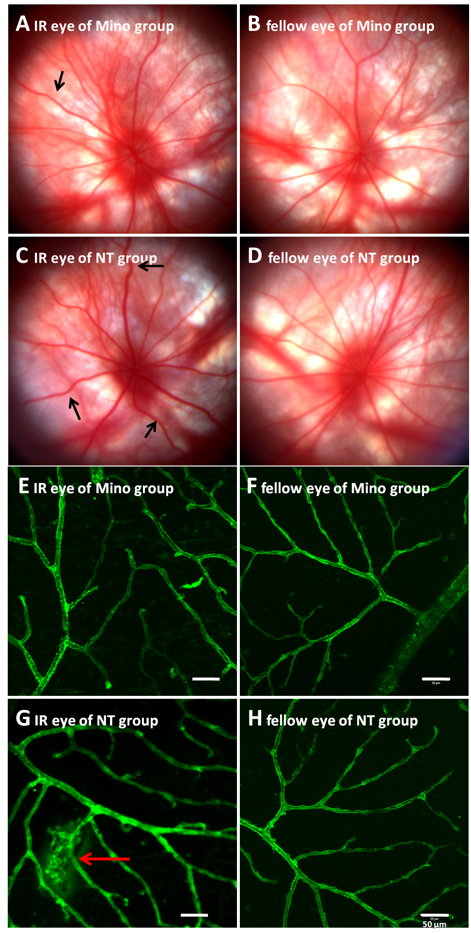
 Figure 2 of
Li, Mol Vis 2021; 27:438-456.
Figure 2 of
Li, Mol Vis 2021; 27:438-456.