Figure 5. EPHA2 is essential for maintaining cell migration ability. A, B: Transwell assays, EPHA2 knockdown reduced cell migration. KD-NC: knockdown using the negative control siRNA, KD-EPHA2: knockdown
of EPHA2, KD-EPHA2+OE-M3: knockdown of EPHA2 + overexpression of M3-EPHA2, KD-EPHA2+OE-WT: knockdown of EPHA2 + overexpression
of WT-EPHA2. C, WT-EPHA2 overexpression restored cell migration ability to a normal level (A). D: M3-EPHA2 overexpression did not rescue the reduced cell migration ability of the EPHA2 kDa cells . E: Quantification of the transwell assay images; five fields were counted in each insert well, n = 5, ***p<0.001, **p<0.01.
F: Western blot images showing that the level of β-catenin significantly decreased in LECs overexpressing M3-EPHA2. G: Quantification of the protein levels based on western blots in F. The relative band intensity was calculated in two steps. First, the values corresponding to the intensity of the β-catenin
band or the FLAG band were divided by those corresponding to the intensity of the β-actin band, and second, these values were
divided by the values obtained for the WT sample. For the WT, this value was set as 1. Endogenous control: β-actin, sample
control: WT-EPHA2 overexpressing group, n = 3, ***p<0.001.
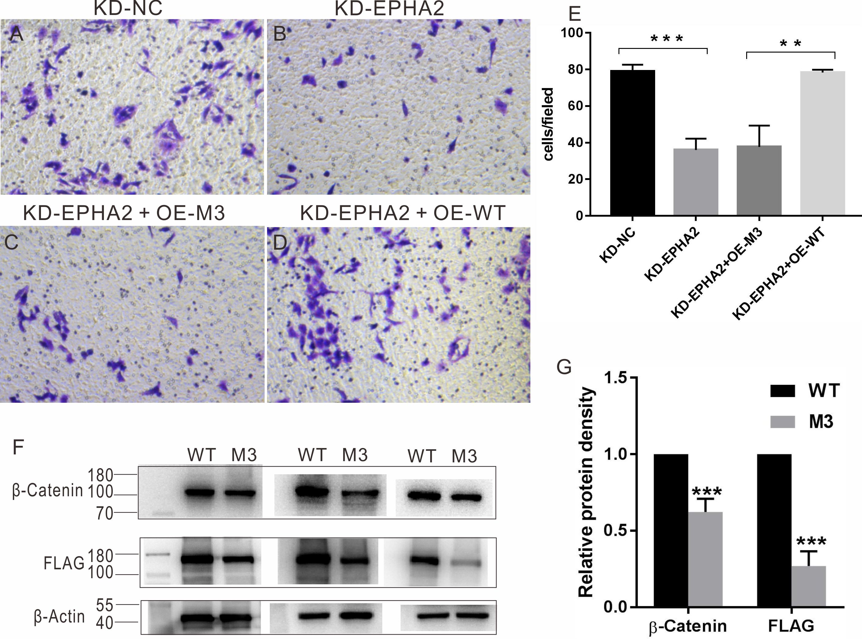
 Figure 5 of
Li, Mol Vis 2021; 27:403-414.
Figure 5 of
Li, Mol Vis 2021; 27:403-414.