 Appendix 3 of
Li, Mol Vis 2021; 27:403-414.
Appendix 3 of
Li, Mol Vis 2021; 27:403-414. Appendix 3 of
Li, Mol Vis 2021; 27:403-414.
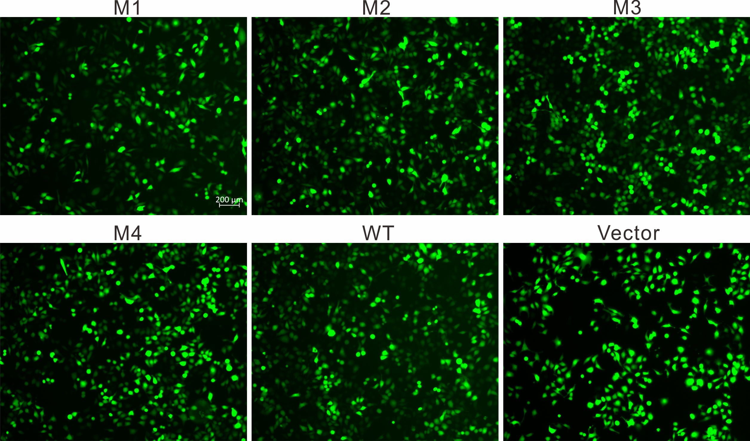
Appendix 3. Fluorescent microscopy indicated the transfection efficiency
To access the data, click or select the words “Appendix 3.” The green fluorescent images of the cells overexpressing WT, M1-M4-EPHA2 and vector plasmid only (as the constructs contain EGFP). Scale bar, 200 µm.