Figure 4. Sibling 2. Full-field electroretinography tracings (upper scotopic, bottom photopic) show normal scotopic (rod) function and
mildly decreased photopic amplitudes (cone function). Rectangular blocks outline the upper and lower limits of normal values.
Stimuli (DA, dark-adopted; LA, light-adapted) were as follows: upper left DA flash 0.01 cd·s·m2, upper right DA flash 3.0 cd·s·m2, lower left LA flash 3.0 cd·s·m2, and lower right LA 0.0 cd·s·m2, flicker at 30 Hz.
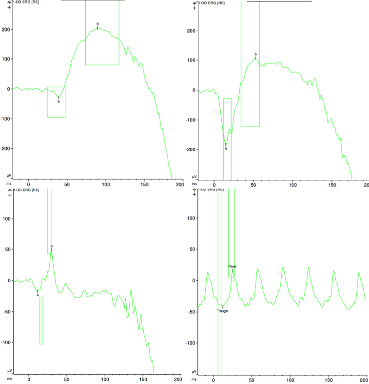
 Figure 4 of
Alsalamah, Mol Vis 2021; 27:396-402.
Figure 4 of
Alsalamah, Mol Vis 2021; 27:396-402.