Figure 2. Sibling 1. A: The appearance of the right eye retina is normal. B: The slit-lamp examination of the right eye shows fine white opacities; these were in both eyes and not visually significant.
C: The appearance of the left eye retina is normal. D, E: Macular spectral-domain optical coherence tomography (SD-OCT) of the right and left eyes reveals subtle findings, which
are shown with magnification in G: The hyporeflective band (outer segments; OS) normally seen in the parafoveal area between the hyper-reflective layers associated
with the inner/outer segment junction (IS/OS) and the RPE/Bruch’s membrane (RPE/BM) complex is not seen; in addition, the
RPE/BM layer has a blurred rather than a sharp appearance. F: Short-wave autofluorescence of the right eye is normal. G: Enlargement of right eye SD-OCT with control. H: Short-wave autofluorescence of the left eye is normal.
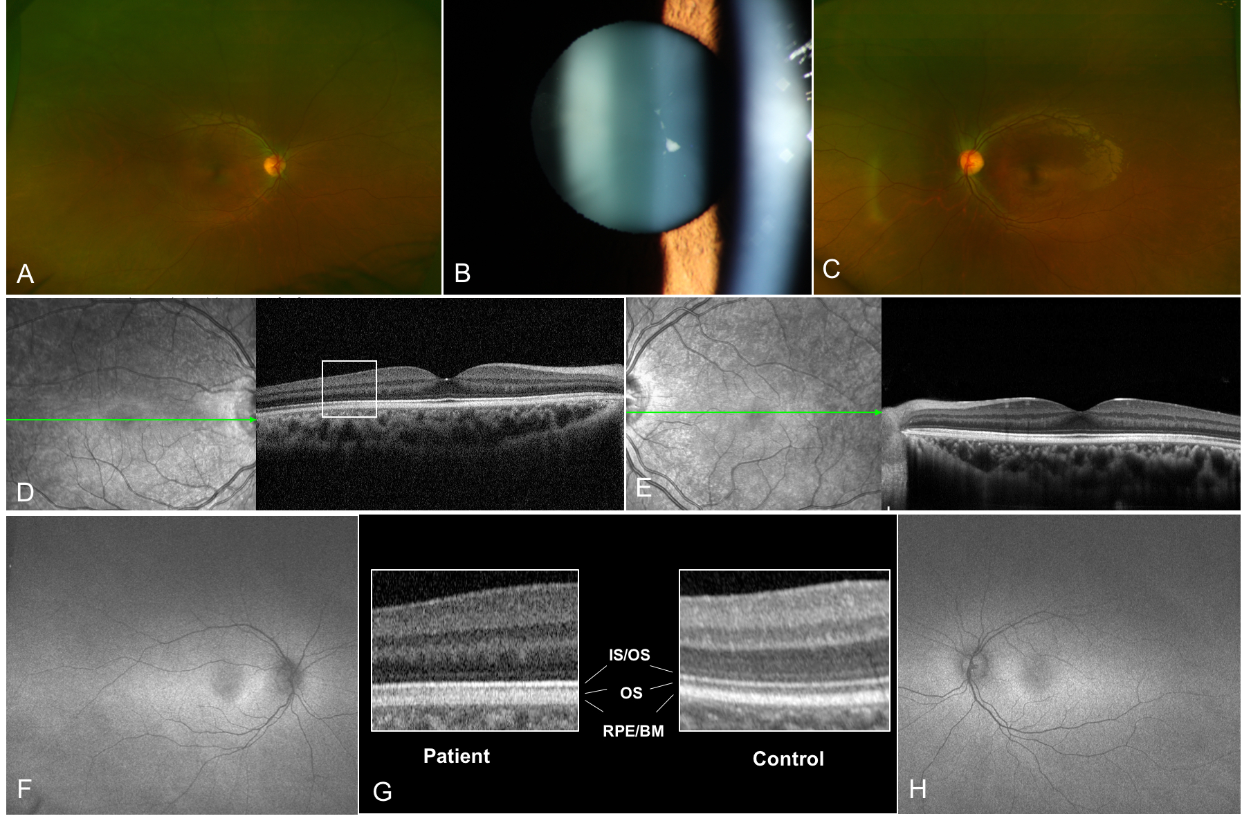
 Figure 2 of
Alsalamah, Mol Vis 2021; 27:396-402.
Figure 2 of
Alsalamah, Mol Vis 2021; 27:396-402.