Figure 3. Expression of Piezo2 in mouse iridocorneal angle tissues. A: Expression of Piezo2 in iridocorneal angle tissues of Piezo2fl/fl mice (n = 5) was determined with western blotting and normalized to the amount of β-actin. Piezo2 was abundant in the mouse
iridocorneal angle tissues compared to Piezo2 expression in the dorsal root ganglia (DRG). Normalized band intensities of
Piezo2 in the DRG and the trabecular meshwork (TM) are displayed. B: Type IV collagen in mouse iridocorneal angle tissues is highlighted with the Collagen IV (ColIV) antibody in green. Piezo2
(red) expression was detected in the TM, the ciliary body’s epithelium, ciliary muscle, and cornea. The nuclei were stained
with 4′,6-diamidino-2-phenylindole (DAPI) in blue. Scale bar: 100 µm.
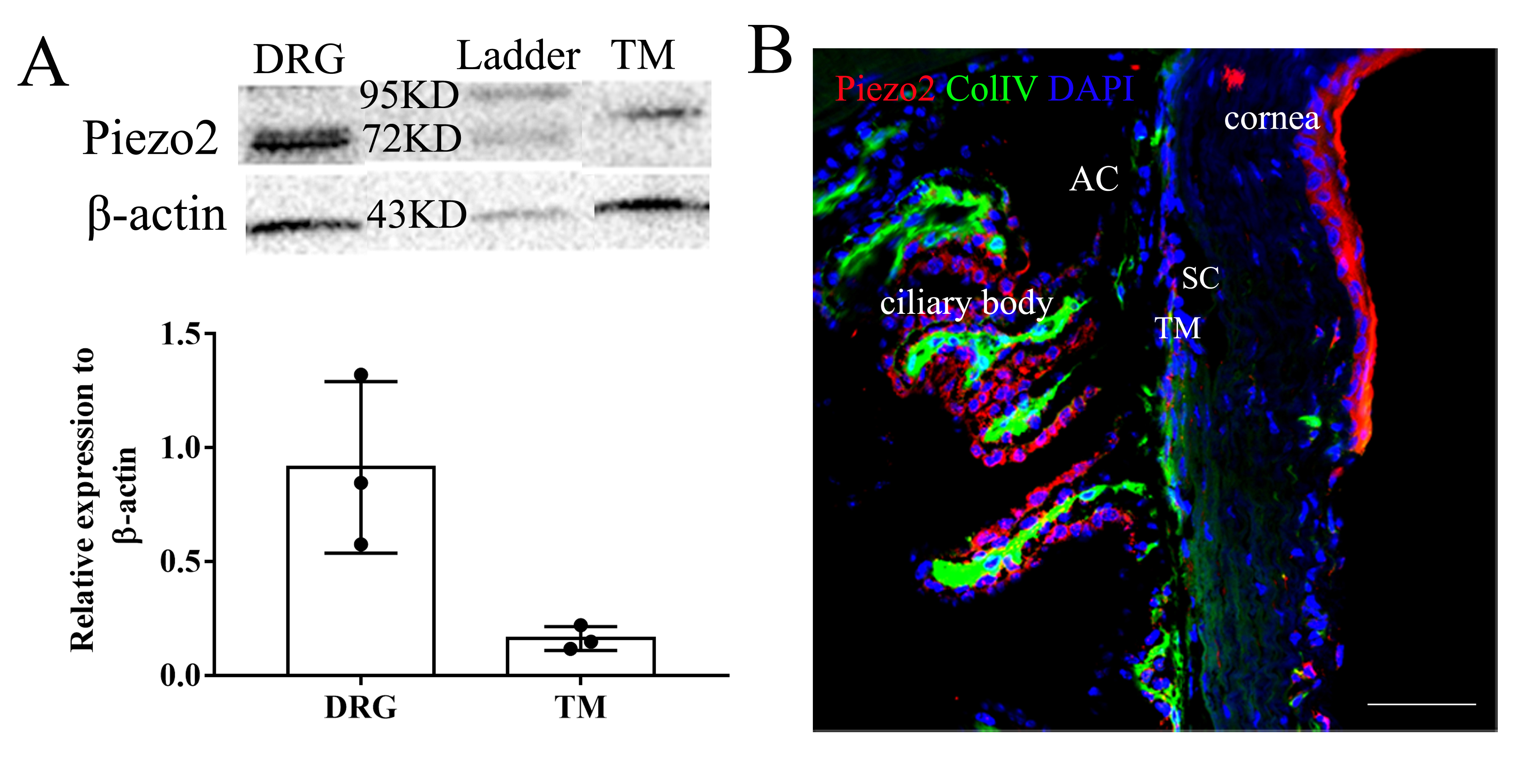
 Figure 3 of
Fang, Mol Vis 2020; 27:354-364.
Figure 3 of
Fang, Mol Vis 2020; 27:354-364.